Wang Shang, Syed Riana, Grishina Olga A, Larina Irina V
Department of Molecular Physiology and Biophysics, Baylor College of Medicine, Houston, Texas.
Department of Bioengineering, Rice University, Houston, Texas.
J Biophotonics. 2018 May;11(5):e201700316. doi: 10.1002/jbio.201700316. Epub 2018 Feb 8.
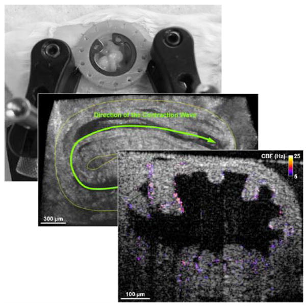
The oviduct (or fallopian tube) serves as an environment for gamete transport, fertilization and preimplantation embryo development in mammals. Although there has been increasing evidence linking infertility with disrupted oviduct function, the specific roles that the oviduct plays in both normal and impaired reproductive processes remain unclear. The mouse is an important mammalian model to study human reproduction. However, most of the current analyses of the mouse oviduct rely on static histology or 2D visualization, and are unable to provide dynamic and volumetric characterization of this organ. The lack of imaging access prevents longitudinal live analysis of the oviduct and its associated reproductive events, limiting the understanding of mechanistic aspects of fertilization and preimplantation pregnancy. To address this limitation, we report a 3D imaging approach that enables prolonged functional assessment of the mouse oviduct in vivo. By combining optical coherence tomography with a dorsal imaging window, this method allows for extended volumetric visualization of the oviduct dynamics, which was previously not achievable. The approach is used for quantitative analysis of oviduct contraction, spatiotemporal characterization of cilia beat frequency and longitudinal imaging. This new approach is a useful in vivo imaging platform for a variety of live studies in mammalian reproduction.
输卵管在哺乳动物中是配子运输、受精和植入前胚胎发育的场所。尽管越来越多的证据表明不孕症与输卵管功能紊乱有关,但输卵管在正常和受损生殖过程中所起的具体作用仍不清楚。小鼠是研究人类生殖的重要哺乳动物模型。然而,目前对小鼠输卵管的大多数分析依赖于静态组织学或二维可视化,无法提供该器官的动态和体积特征。缺乏成像途径阻碍了对输卵管及其相关生殖事件的纵向实时分析,限制了对受精和植入前妊娠机制方面的理解。为了解决这一限制,我们报告了一种三维成像方法,该方法能够在体内对小鼠输卵管进行长期功能评估。通过将光学相干断层扫描与背部成像窗口相结合,这种方法可以对输卵管动态进行扩展的体积可视化,这在以前是无法实现的。该方法用于输卵管收缩的定量分析、纤毛搏动频率的时空特征分析和纵向成像。这种新方法是一个有用的体内成像平台,可用于哺乳动物生殖的各种实时研究。