Department of Methods and Experimental Psychology, Faculty of Psychology and Education, University of Deusto, Bilbao, Basque Country, Spain.
Research Imaging Centre, Campbell Family Mental Health Research Institute, Centre for Addiction and Mental Health, University of Toronto, Toronto, Ontario, Canada; Division of Brain, Imaging and Behaviour - Systems Neuroscience, Krembil Research Institute, UHN, University of Toronto, Ontario, Canada.
Neuroimage Clin. 2017 Dec 9;17:847-855. doi: 10.1016/j.nicl.2017.12.013. eCollection 2018.
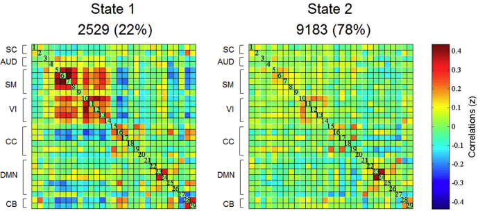
The objective was to assess dynamic functional connectivity (FC) and local/global connectivity in Parkinson's disease (PD) patients with mild cognitive impairment (PD-MCI) and with normal cognition (PD-NC). The sample included 35 PD patients and 26 healthy controls (HC). Cognitive assessment followed an extensive neuropsychological battery. For resting-state functional MRI (rs-fMRI) analysis, independent component analysis (ICA) was performed and components were located in 7 networks: Subcortical (SC), Auditory (AUD), Somatomotor (SM), visual (VI), cognitive-control (CC), default-mode (DMN), and cerebellar (CB). Dynamic FC analysis was performed using the GIFT toolbox. FC differences between groups in each FC state were analysed with the network-based statistic (NBS) approach. Finally, a graph-theoretical analysis for local/global parameters was performed. The whole sample showed 2 dynamic FC states during the rs-fMRI. PD-MCI patients showed decreased mean dwell time in the hypo-connectivity state ( = 0.030) and showed increased number of state transitions ( = 0.007) compared with the HC. In addition, in the hypo-connectivity state, PD-MCI patients showed reduced inter-network FC between the SM-CC, SM-VI, SM-AUD, CC-VI and SC-DMN compared with the HC ( < 0.05-FDR). These FC alterations in PD-MCI were accompanied by graph-topological alterations in nodes located in the SM network ( < 0.001). In contrast, no differences were found between the PD-NC and HC. Findings suggest the presence of dynamic functional brain deteriorations in PD-MCI that are not present in PD-NC, showing the PD-MCI group dynamic FC dysfunctions, reduced FC mostly between SM-CC networks and graph-topological deteriorations in the SM network. A dynamic FC approach could be helpful to understand cognitive deterioration in PD.
目的是评估轻度认知障碍(PD-MCI)和认知正常(PD-NC)的帕金森病(PD)患者的动态功能连接(FC)和局部/全局连接。该样本包括 35 名 PD 患者和 26 名健康对照(HC)。认知评估遵循广泛的神经心理学测试。对于静息态功能磁共振成像(rs-fMRI)分析,进行了独立成分分析(ICA),并将成分定位在 7 个网络中:皮质下(SC)、听觉(AUD)、运动(SM)、视觉(VI)、认知控制(CC)、默认模式(DMN)和小脑(CB)。使用 GIFT 工具箱进行动态 FC 分析。使用基于网络的统计(NBS)方法分析每个 FC 状态下组间的 FC 差异。最后,进行了局部/全局参数的图论分析。整个样本在 rs-fMRI 期间显示出 2 种动态 FC 状态。与 HC 相比,PD-MCI 患者在低连接状态下的平均停留时间减少(=0.030),状态转换次数增加(=0.007)。此外,在低连接状态下,PD-MCI 患者与 HC 相比,SM-CC、SM-VI、SM-AUD、CC-VI 和 SC-DMN 之间的网络间 FC 减少(<0.05-FDR)。在 PD-MCI 中,这些 FC 改变伴随着位于 SM 网络中的节点的图拓扑改变(<0.001)。相比之下,PD-NC 和 HC 之间没有差异。研究结果表明,PD-MCI 存在动态功能脑恶化,而 PD-NC 则不存在,表明 PD-MCI 组存在动态 FC 功能障碍,SM-CC 网络之间的 FC 减少,SM 网络的图拓扑恶化。动态 FC 方法可能有助于理解 PD 中的认知恶化。