• 文献检索
  • 文档翻译
  • 深度研究
  • 学术资讯
  • Suppr Zotero 插件Zotero 插件
  • 邀请有礼
  • 套餐&价格
  • 历史记录
应用&插件
Suppr Zotero 插件Zotero 插件浏览器插件Mac 客户端Windows 客户端微信小程序
定价
高级版会员购买积分包购买API积分包
服务
文献检索文档翻译深度研究API 文档MCP 服务
关于我们
关于 Suppr公司介绍联系我们用户协议隐私条款
关注我们

Suppr 超能文献

核心技术专利:CN118964589B侵权必究
粤ICP备2023148730 号-1Suppr @ 2026

文献检索

告别复杂PubMed语法,用中文像聊天一样搜索,搜遍4000万医学文献。AI智能推荐,让科研检索更轻松。

立即免费搜索

文件翻译

保留排版,准确专业,支持PDF/Word/PPT等文件格式,支持 12+语言互译。

免费翻译文档

深度研究

AI帮你快速写综述,25分钟生成高质量综述,智能提取关键信息,辅助科研写作。

立即免费体验

常规制作的小鼠脑切片图像的几何处理。

Geometry processing of conventionally produced mouse brain slice images.

机构信息

Department of Computer Science, University of California, Irvine, CA 92697-3435, United States.

Department of Anatomy and Neurobiology, School of Medicine, University of California, Irvine, CA 92697-1275, United States; Department of Biomedical Engineering, University of California, Irvine, CA 92697-2715, United States; Department of Electrical Engineering and Computer Science, University of California, Irvine, CA 92697-2625, United States.

出版信息

J Neurosci Methods. 2018 Aug 1;306:45-56. doi: 10.1016/j.jneumeth.2018.04.008. Epub 2018 Apr 22.

DOI:10.1016/j.jneumeth.2018.04.008
PMID:29689283
原文链接:https://pmc.ncbi.nlm.nih.gov/articles/PMC6086593/
Abstract

BACKGROUND

Brain mapping research in most neuroanatomical laboratories relies on conventional processing techniques, which often introduce histological artifacts such as tissue tears and tissue loss.

NEW METHOD

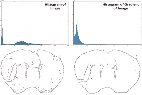
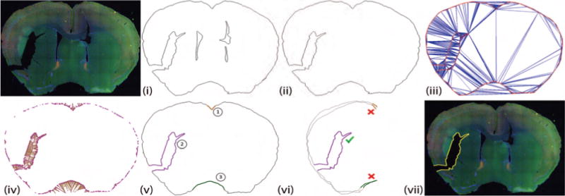
In this paper, we present techniques and algorithms for automatic registration and 3D reconstruction of conventionally produced mouse brain slices in a standardized atlas space. This is achieved first by constructing a virtual 3D mouse brain model from annotated slices of Allen Reference Atlas (ARA). Virtual re-slicing of the reconstructed model generates ARA-based slice images corresponding to the microscopic images of histological brain sections. These image pairs are aligned using a geometric approach through contour images. Histological artifacts in the microscopic images are detected and removed using Constrained Delaunay Triangulation before performing global alignment. Finally, non-linear registration is performed by solving Laplace's equation with Dirichlet boundary conditions.

RESULTS

Our methods provide significant improvements over previously reported registration techniques for the tested slices in 3D space, especially on slices with significant histological artifacts. Further, as one of the application we count the number of neurons in various anatomical regions using a dataset of 51 microscopic slices from a single mouse brain.

COMPARISON WITH EXISTING METHOD(S): To the best of our knowledge the presented work is the first that automatically registers both clean as well as highly damaged high-resolutions histological slices of mouse brain to a 3D annotated reference atlas space.

CONCLUSIONS

This work represents a significant contribution to this subfield of neuroscience as it provides tools to neuroanatomist for analyzing and processing histological data.

摘要

背景

大多数神经解剖学实验室的脑图谱研究依赖于传统的处理技术,这些技术往往会引入组织撕裂和组织丢失等组织学伪影。

新方法

在本文中,我们提出了一种用于自动注册和以标准化图谱空间对常规制作的小鼠脑切片进行 3D 重建的技术和算法。这首先是通过从艾伦参考图谱(ARA)的注释切片构建虚拟 3D 小鼠脑模型来实现的。重建模型的虚拟重新切片生成与组织学脑切片的显微镜图像相对应的基于 ARA 的切片图像。通过轮廓图像使用几何方法对这些图像对进行对齐。在执行全局对齐之前,使用约束 Delaunay 三角剖分检测并去除显微镜图像中的组织学伪影。最后,通过求解具有狄利克雷边界条件的拉普拉斯方程来进行非线性注册。

结果

我们的方法在 3D 空间中对测试切片的注册技术提供了显著的改进,尤其是在具有明显组织学伪影的切片上。此外,作为应用之一,我们使用来自单个小鼠大脑的 51 个微观切片数据集来计算各种解剖区域的神经元数量。

与现有方法的比较

据我们所知,目前的工作是第一个自动将清洁和高度损坏的高分辨率组织学小鼠脑切片注册到 3D 注释参考图谱空间的工作。

结论

这项工作代表了神经科学这一分支的重要贡献,因为它为神经解剖学家提供了分析和处理组织学数据的工具。

https://cdn.ncbi.nlm.nih.gov/pmc/blobs/6da6/6086593/cb8347d5a6d0/nihms977652f11.jpg
https://cdn.ncbi.nlm.nih.gov/pmc/blobs/6da6/6086593/2a8cdd050d95/nihms977652f1.jpg
https://cdn.ncbi.nlm.nih.gov/pmc/blobs/6da6/6086593/ce0103d74099/nihms977652f2.jpg
https://cdn.ncbi.nlm.nih.gov/pmc/blobs/6da6/6086593/03bac2fa5eea/nihms977652f3.jpg
https://cdn.ncbi.nlm.nih.gov/pmc/blobs/6da6/6086593/470753212739/nihms977652f4.jpg
https://cdn.ncbi.nlm.nih.gov/pmc/blobs/6da6/6086593/155203acf47a/nihms977652f5.jpg
https://cdn.ncbi.nlm.nih.gov/pmc/blobs/6da6/6086593/5faa2826d216/nihms977652f6.jpg
https://cdn.ncbi.nlm.nih.gov/pmc/blobs/6da6/6086593/19b2a837b95d/nihms977652f7.jpg
https://cdn.ncbi.nlm.nih.gov/pmc/blobs/6da6/6086593/218d6c46a392/nihms977652f8.jpg
https://cdn.ncbi.nlm.nih.gov/pmc/blobs/6da6/6086593/7c5313e25469/nihms977652f9.jpg
https://cdn.ncbi.nlm.nih.gov/pmc/blobs/6da6/6086593/eb00d5ef1803/nihms977652f10.jpg
https://cdn.ncbi.nlm.nih.gov/pmc/blobs/6da6/6086593/cb8347d5a6d0/nihms977652f11.jpg

相似文献

1
Geometry processing of conventionally produced mouse brain slice images.常规制作的小鼠脑切片图像的几何处理。
J Neurosci Methods. 2018 Aug 1;306:45-56. doi: 10.1016/j.jneumeth.2018.04.008. Epub 2018 Apr 22.
2
Localization and Registration of 2D Histological Mouse Brain Images in 3D Atlas Space.二维组织学小鼠脑图像在三维图谱空间中的定位和配准。
Neuroinformatics. 2023 Jul;21(3):615-630. doi: 10.1007/s12021-023-09632-8. Epub 2023 Jun 26.
3
Constructing and optimizing 3D atlases from 2D data with application to the developing mouse brain.从二维数据构建和优化 3D 图谱及其在发育中老鼠大脑中的应用。
Elife. 2021 Feb 11;10:e61408. doi: 10.7554/eLife.61408.
4
Precise Mapping of Single Neurons by Calibrated 3D Reconstruction of Brain Slices Reveals Topographic Projection in Mouse Visual Cortex.通过校准的脑片 3D 重建对单个神经元进行精确映射,揭示了小鼠视觉皮层中的拓扑投影。
Cell Rep. 2020 May 26;31(8):107682. doi: 10.1016/j.celrep.2020.107682.
5
Automatic navigation system for the mouse brain.鼠标脑自动导航系统。
J Comp Neurol. 2019 Sep 1;527(13):2200-2211. doi: 10.1002/cne.24635. Epub 2019 Feb 24.
6
Mapping Histological Slice Sequences to the Allen Mouse Brain Atlas Without 3D Reconstruction.无需三维重建将组织学切片序列映射到艾伦小鼠脑图谱
Front Neuroinform. 2018 Dec 11;12:93. doi: 10.3389/fninf.2018.00093. eCollection 2018.
7
Development of a 2D Image Reconstruction and Viewing System for Histological Images from Multiple Tissue Blocks: Towards High-Resolution Whole-Organ 3D Histological Images.用于多个组织块组织学图像的二维图像重建与查看系统的开发:迈向高分辨率全器官三维组织学图像
Pathobiology. 2016;83(2-3):127-39. doi: 10.1159/000443278. Epub 2016 Apr 26.
8
MRI-guided volume reconstruction of mouse brain from histological sections.MRI 引导的鼠脑组织切片容积重建
J Neurosci Methods. 2012 Nov 15;211(2):210-7. doi: 10.1016/j.jneumeth.2012.08.021. Epub 2012 Sep 7.
9
Robust joint registration of multiple stains and MRI for multimodal 3D histology reconstruction: Application to the Allen human brain atlas.多模态 3D 组织学重建的多染色和 MRI 的稳健联合配准:Allen 人类大脑图谱的应用。
Med Image Anal. 2022 Jan;75:102265. doi: 10.1016/j.media.2021.102265. Epub 2021 Oct 16.
10
Automatic best reference slice selection for smooth volume reconstruction of a mouse brain from histological images.自动选择最佳参考切片,以便从组织学图像中对小鼠大脑进行平滑体积重建。
IEEE Trans Med Imaging. 2010 Sep;29(9):1688-96. doi: 10.1109/TMI.2010.2050594. Epub 2010 Jun 14.

引用本文的文献

1
SlicesMapi: An Interactive Three-Dimensional Registration Method for Serial Histological Brain Slices.切片映射:一种用于连续组织学脑切片的交互式三维配准方法。
Neuroinformatics. 2025 Apr 16;23(2):28. doi: 10.1007/s12021-025-09724-7.
2
RegBoost: Enhancing mouse brain image registration using geometric priors and Laplacian interpolation.RegBoost:利用几何先验和拉普拉斯插值增强小鼠脑图像配准
Neuroimage. 2025 Jan;305:120981. doi: 10.1016/j.neuroimage.2024.120981. Epub 2024 Dec 26.
3
Molecular signatures of cortical expansion in the human foetal brain.

本文引用的文献

1
Neuroinformatics of the Allen Mouse Brain Connectivity Atlas.艾伦小鼠脑连接图谱的神经信息学
Methods. 2015 Feb;73:4-17. doi: 10.1016/j.ymeth.2014.12.013. Epub 2014 Dec 20.
2
Whole-brain mapping of behaviourally induced neural activation in mice.小鼠行为诱导神经激活的全脑图谱
Brain Struct Funct. 2015 Jul;220(4):2043-57. doi: 10.1007/s00429-014-0774-0. Epub 2014 Apr 24.
3
A mesoscale connectome of the mouse brain.小鼠大脑的介观连接组图谱
人类胎儿大脑皮层扩张的分子特征。
Nat Commun. 2024 Nov 8;15(1):9685. doi: 10.1038/s41467-024-54034-2.
4
Fine-tuning TrailMap: The utility of transfer learning to improve the performance of deep learning in axon segmentation of light-sheet microscopy images.微调 TrailMap:迁移学习在提高光片显微镜图像轴突分割中深度学习性能的应用。
PLoS One. 2024 Mar 29;19(3):e0293856. doi: 10.1371/journal.pone.0293856. eCollection 2024.
5
Molecular signatures of cortical expansion in the human fetal brain.人类胎儿大脑皮质扩张的分子特征
bioRxiv. 2024 Feb 13:2024.02.13.580198. doi: 10.1101/2024.02.13.580198.
6
Automated identification of protein expression intensity and classification of protein cellular locations in mouse brain regions from immunofluorescence images.从免疫荧光图像中自动识别小鼠脑区的蛋白质表达强度和蛋白质细胞定位分类。
Med Biol Eng Comput. 2024 Apr;62(4):1105-1119. doi: 10.1007/s11517-023-02985-x. Epub 2023 Dec 27.
7
Fine-tuning TrailMap: The utility of transfer learning to improve the performance of deep learning in axon segmentation of light-sheet microscopy images.微调轨迹图:迁移学习在提高深度学习对光片显微镜图像轴突分割性能方面的效用。
bioRxiv. 2023 Oct 23:2023.10.23.563546. doi: 10.1101/2023.10.23.563546.
8
Expected affine: A registration method for damaged section in serial sections electron microscopy.预期仿射变换:一种用于连续切片电子显微镜中受损切片的配准方法。
Front Neuroinform. 2022 Sep 2;16:944050. doi: 10.3389/fninf.2022.944050. eCollection 2022.
9
BoutonNet: an automatic method to detect anterogradely labeled presynaptic boutons in brain tissue sections.BoutonNet:一种自动检测脑组织切片中顺向标记的突触前末梢的方法。
Brain Struct Funct. 2022 Jul;227(6):1921-1932. doi: 10.1007/s00429-022-02504-y. Epub 2022 Jun 1.
10
ARMBIS: accurate and robust matching of brain image sequences from multiple modal imaging techniques.ARMBIS:来自多种模态成像技术的大脑图像序列的精确和稳健匹配。
Bioinformatics. 2019 Dec 15;35(24):5281-5289. doi: 10.1093/bioinformatics/btz404.
Nature. 2014 Apr 10;508(7495):207-14. doi: 10.1038/nature13186. Epub 2014 Apr 2.
4
Cell-type-specific circuit connectivity of hippocampal CA1 revealed through Cre-dependent rabies tracing.通过Cre依赖的狂犬病追踪揭示海马CA1区细胞类型特异性的回路连接性。
Cell Rep. 2014 Apr 10;7(1):269-80. doi: 10.1016/j.celrep.2014.02.030. Epub 2014 Mar 20.
5
Similarity-based appearance-prior for fitting a subdivision mesh in gene expression images.基于相似度的外观先验,用于在基因表达图像中拟合细分网格。
Med Image Comput Comput Assist Interv. 2012;15(Pt 1):577-84. doi: 10.1007/978-3-642-33415-3_71.
6
The structure of the nervous system of the nematode Caenorhabditis elegans.秀丽隐杆线虫的神经系统结构。
Philos Trans R Soc Lond B Biol Sci. 1986 Nov 12;314(1165):1-340. doi: 10.1098/rstb.1986.0056.
7
Serial two-photon tomography for automated ex vivo mouse brain imaging.用于自动离体鼠脑成像的串行双光子断层扫描。
Nat Methods. 2012 Jan 15;9(3):255-8. doi: 10.1038/nmeth.1854.
8
The future of the human connectome.人类连接组计划的未来。
Neuroimage. 2012 Aug 15;62(2):1299-310. doi: 10.1016/j.neuroimage.2012.01.032. Epub 2012 Jan 10.
9
The big and the small: challenges of imaging the brain's circuits.大与小:脑回路成像的挑战。
Science. 2011 Nov 4;334(6056):618-23. doi: 10.1126/science.1209168.
10
Three-dimensional reconstruction of serial mouse brain sections: solution for flattening high-resolution large-scale mosaics.三维重建连续的小鼠脑切片:用于平铺高分辨率大尺度镶嵌图的解决方案。
Front Neuroanat. 2011 Mar 7;5:17. doi: 10.3389/fnana.2011.00017. eCollection 2011.