Feizi Sepehr, Delfazayebaher Siamak, Javadi Mohammad Ali, Karimian Farid, Ownagh Vahid, Sadeghpour Fatemeh
Ocular Tissue Engineering Research Center, Shahid Beheshti University of Medical Sciences, Tehran, Iran.
Ophthalmic Research Center, Shahid Beheshti University of Medical Sciences, Tehran, Iran.
J Ophthalmic Vis Res. 2018 Apr-Jun;13(2):93-100. doi: 10.4103/jovr.jovr_19_17.
To compare mean posterior corneal power and astigmatism in normal versus keratoconus affected eyes and determine the optimal cut-off points to maximize sensitivity and specificity in discriminating keratoconus from normal corneas.
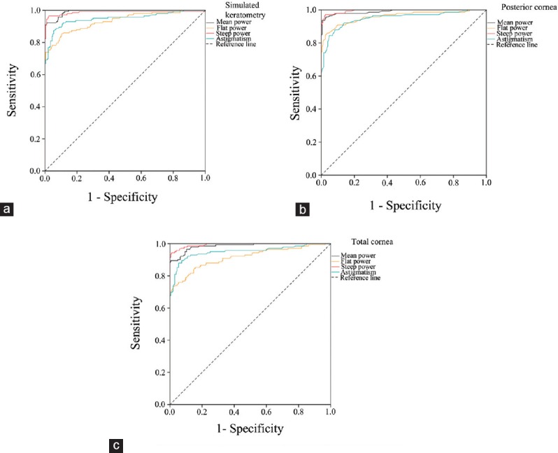
A total of 204 normal eyes and 142 keratoconus affected eyes were enrolled in this prospective comparative study. Mean posterior corneal power and astigmatism were measured using a dual Scheimpflug camera. Correlation coefficients were calculated to assess the relationship between the magnitudes of keratometric and posterior corneal astigmatism in the study groups. Receiver operating characteristic curves were used to compare the sensitivity and specificity of the measured parameters and to identify the optimal cut-off points for discriminating keratoconus from normal corneas.
The mean posterior corneal power was -6.29 ± 0.20 D in the normal group and -7.77 ± 0.87 D in the keratoconus group ( < 0.001). The mean magnitudes of the posterior corneal astigmatisms were -0.32 ± 0.15 D and -0.94 ± 0.39 D in the normal and keratoconus groups, respectively ( < 0.001). Significant correlations were found between the magnitudes of keratometric and posterior corneal astigmatism in the normal (r=-0.76, < 0.001) and keratoconus (r=-0.72, < 0.001) groups. The mean posterior corneal power and astigmatism were highly reliable characteristics that distinguished keratoconus from normal corneas (area under the curve, 0.99 and 0.95, respectively). The optimal cut-off points of mean posterior corneal power and astigmatism were -6.70 D and -0.54 D, respectively.
Mean posterior corneal power and astigmatism measured using a Galilei analyzer camera might have potential in diagnosing keratoconus. The cut-off points provided can be used for keratoconus screening.
比较正常眼与圆锥角膜患眼的角膜后表面平均屈光力和散光情况,并确定在区分圆锥角膜与正常角膜时使敏感度和特异度最大化的最佳截断点。
本前瞻性对照研究共纳入204只正常眼和142只圆锥角膜患眼。使用双Scheimpflug相机测量角膜后表面平均屈光力和散光。计算相关系数以评估研究组中角膜曲率计测量的散光与角膜后表面散光大小之间的关系。使用受试者工作特征曲线比较测量参数的敏感度和特异度,并确定区分圆锥角膜与正常角膜的最佳截断点。
正常组角膜后表面平均屈光力为-6.29±0.20D,圆锥角膜组为-7.77±0.87D(P<0.001)。正常组和圆锥角膜组角膜后表面散光的平均大小分别为-0.32±0.15D和-0.94±0.39D(P<0.001)。正常组(r=-0.76,P<0.001)和圆锥角膜组(r=-0.72,P<0.001)中角膜曲率计测量的散光与角膜后表面散光大小之间存在显著相关性。角膜后表面平均屈光力和散光为区分圆锥角膜与正常角膜的高度可靠特征(曲线下面积分别为0.99和0.95)。角膜后表面平均屈光力和散光的最佳截断点分别为-6.70D和-0.54D。
使用Galilei分析仪相机测量的角膜后表面平均屈光力和散光可能在圆锥角膜诊断中具有潜力。所提供的截断点可用于圆锥角膜筛查。