Kao Chung-Wei, Wu Po-Ting, Liao Mei-Yi, Chung I-Ju, Yang Kai-Chien, Tseng Wen-Yih Isaac, Yu Jiashing
Department of Chemical Engineering, National Taiwan University, Taipei 10617, Taiwan.
Department of Applied Chemistry, National Pingtung University, Pingtung 90003, Taiwan.
Pharmaceutics. 2018 May 24;10(2):62. doi: 10.3390/pharmaceutics10020062.
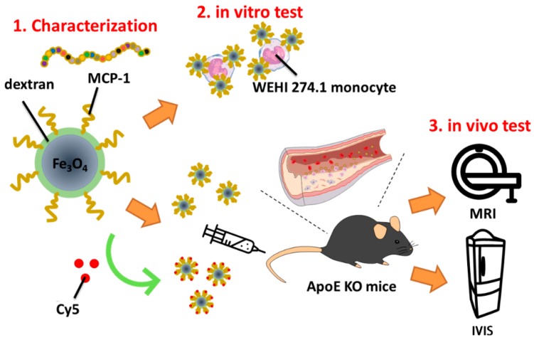
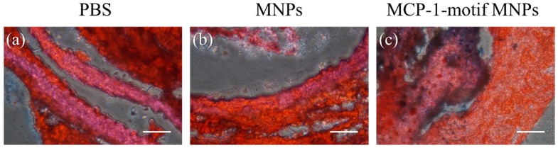
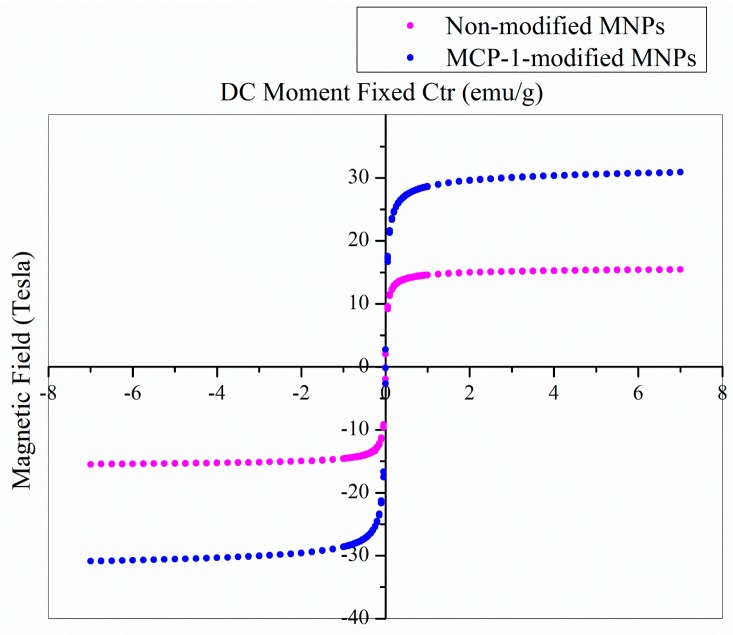
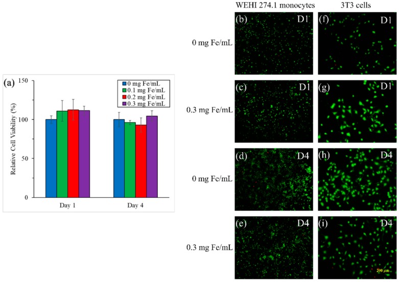
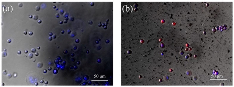
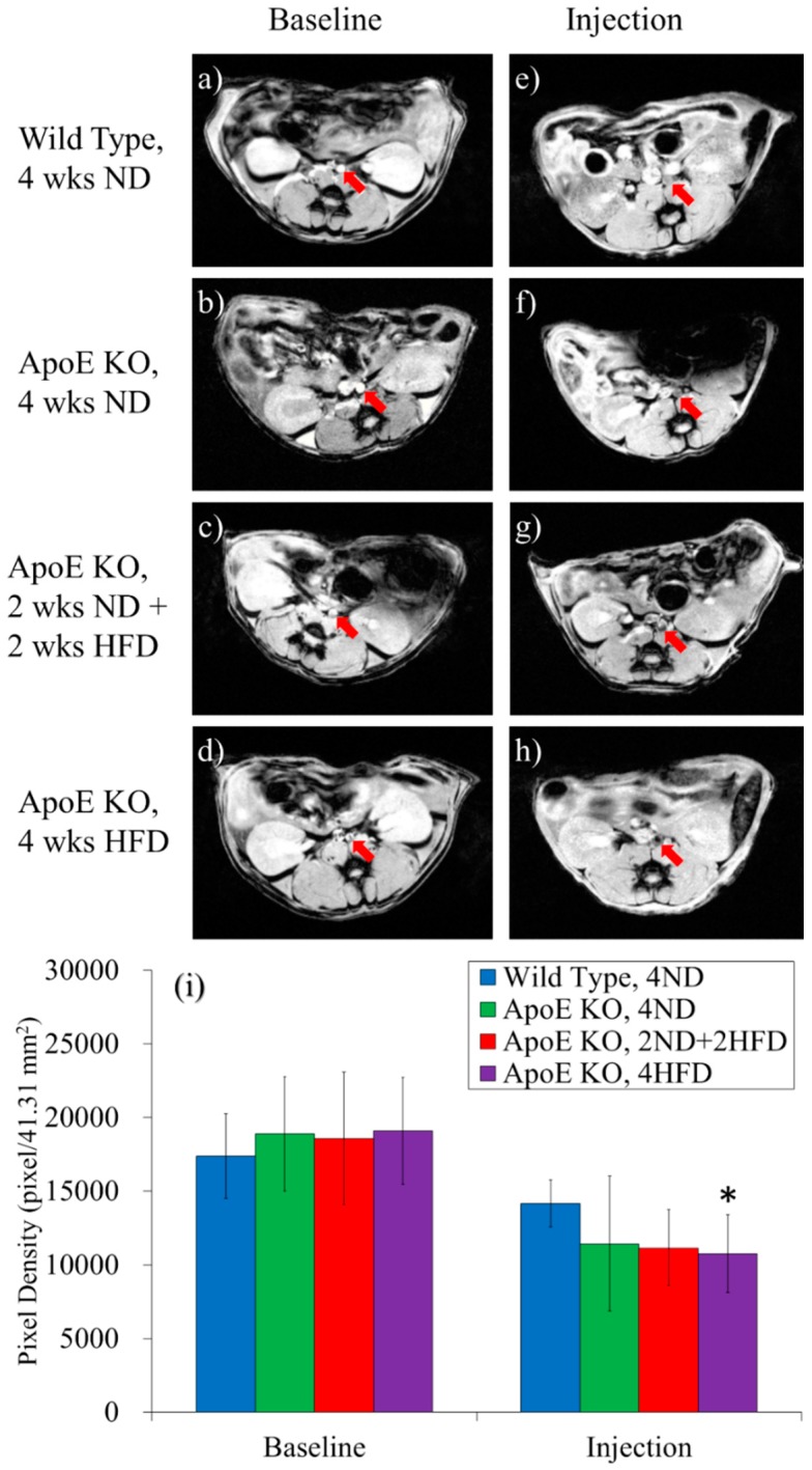
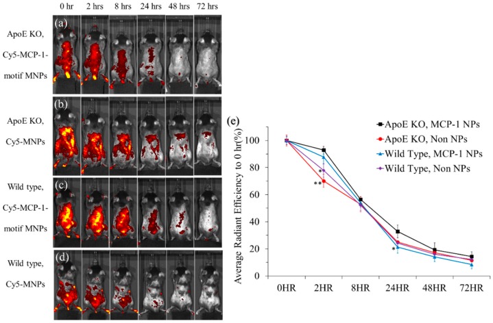
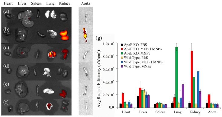
Atherosclerosis is a multifactorial inflammatory disease that may progress silently for long period, and it is also widely accepted as the main cause of cardiovascular diseases. To prevent atherosclerotic plaques from generating, imaging early molecular markers and quantifying the extent of disease progression are desired. During inflammation, circulating monocytes leave the bloodstream and migrate into incipient lipid accumulation in the artery wall, following conditioning by local growth factors and proinflammatory cytokines; therefore, monocyte accumulation in the arterial wall can be observed in fatty streaks, rupture-prone plaques, and experimental atherosclerosis. In this work, we synthesized monocyte-targeting iron oxide magnetic nanoparticles (MNPs), which were incorporated with the peptides derived from the chemokine receptor C-C chemokine receptor type 2 (CCR2)-binding motif of monocytes chemoattractant protein-1 (MCP-1) as a diagnostic tool for potential atherosclerosis. MCP-1-motif MNPs co-localized with monocytes in in vitro fluorescence imaging. In addition, with MNPs injection in ApoE knockout mice (ApoE KO mice), the well-characterized animal model of atherosclerosis, MNPs were found in specific organs or regions which had monocytes accumulation, especially the aorta of atherosclerosis model mice, through in vivo imaging system (IVIS) imaging and magnetic resonance imaging (MRI). We also performed Oil Red O staining and Prussian Blue staining to confirm the co-localization of MCP-1-motif MNPs and atherosclerosis. The results showed the promising potential of MCP-1-motif MNPs as a diagnostic agent of atherosclerosis.
动脉粥样硬化是一种多因素炎症性疾病,可能会长期悄然进展,并且它也被广泛认为是心血管疾病的主要原因。为了预防动脉粥样硬化斑块的形成,人们期望能够成像早期分子标志物并量化疾病进展的程度。在炎症过程中,循环中的单核细胞离开血液,在局部生长因子和促炎细胞因子的作用下,迁移到动脉壁初期脂质积聚处;因此,在脂肪条纹、易破裂斑块和实验性动脉粥样硬化中都可以观察到单核细胞在动脉壁中的积聚。在这项工作中,我们合成了靶向单核细胞的氧化铁磁性纳米颗粒(MNPs),其结合了源自单核细胞趋化蛋白-1(MCP-1)的C-C趋化因子受体2型(CCR2)结合基序的肽,作为潜在动脉粥样硬化的诊断工具。MCP-1基序MNPs在体外荧光成像中与单核细胞共定位。此外,在动脉粥样硬化特征明确的动物模型载脂蛋白E基因敲除小鼠(ApoE KO小鼠)中注射MNPs后,通过体内成像系统(IVIS)成像和磁共振成像(MRI)发现,MNPs存在于有单核细胞积聚的特定器官或区域,尤其是动脉粥样硬化模型小鼠的主动脉。我们还进行了油红O染色和普鲁士蓝染色,以确认MCP-1基序MNPs与动脉粥样硬化的共定位。结果显示MCP-1基序MNPs作为动脉粥样硬化诊断剂具有广阔的应用前景。