Institute of Information Systems and Applications, National Tsing Hua University, Hsinchu, Taiwan.
Department of Computer Science and Information Engineering, National Taichung University of Science and Technology, Taichung, Taiwan.
J Healthc Eng. 2018 Apr 3;2018:2908517. doi: 10.1155/2018/2908517. eCollection 2018.
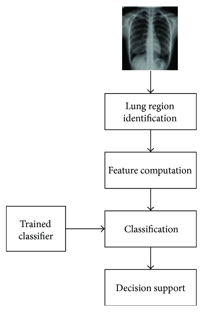
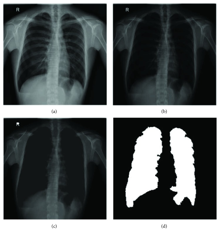
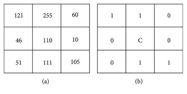
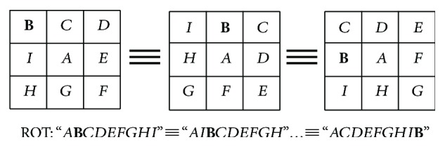
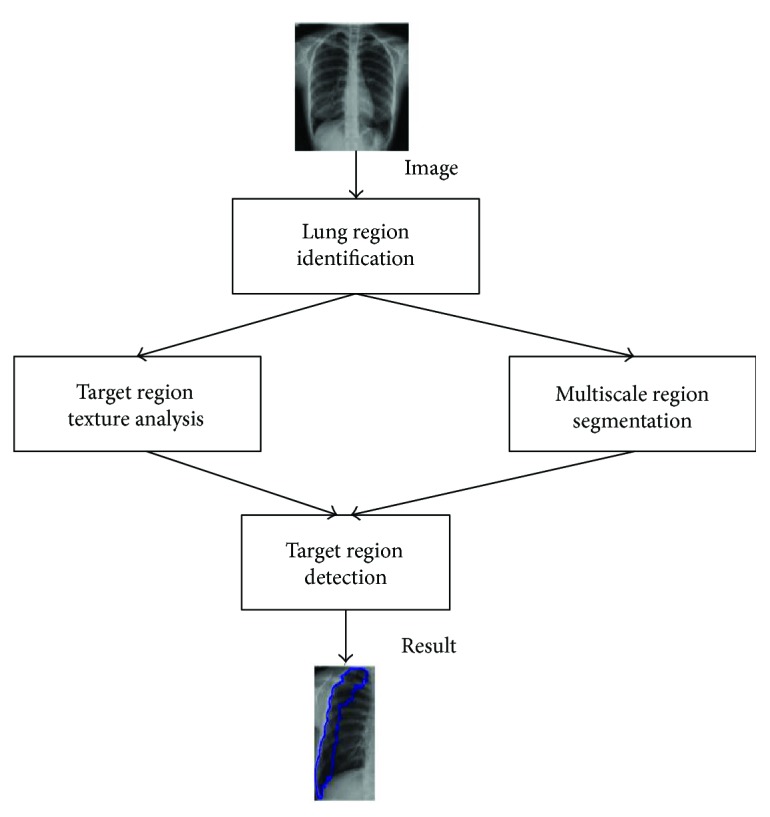
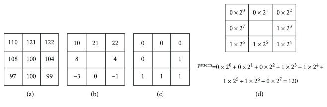
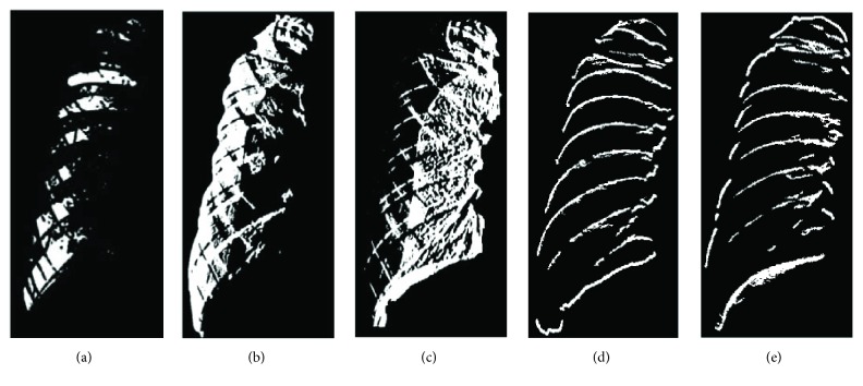
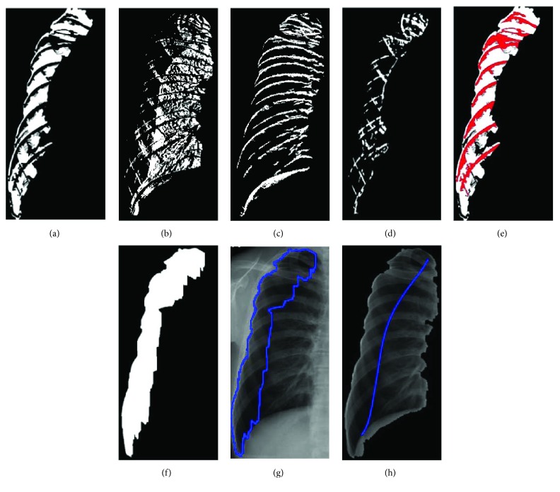
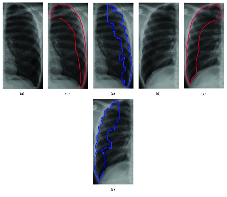
Automatic image segmentation and feature analysis can assist doctors in the treatment and diagnosis of diseases more accurately. Automatic medical image segmentation is difficult due to the varying image quality among equipment. In this paper, the automatic method employed image multiscale intensity texture analysis and segmentation to solve this problem. In this paper, firstly, SVM is applied to identify common pneumothorax. Features are extracted from lung images with the LBP (local binary pattern). Then, classification of pneumothorax is determined by SVM. Secondly, the proposed automatic pneumothorax detection method is based on multiscale intensity texture segmentation by removing the background and noises in chest images for segmenting abnormal lung regions. The segmentation of abnormal regions is used for texture transformed from computing multiple overlapping blocks. The rib boundaries are identified with Sobel edge detection. Finally, in obtaining a complete disease region, the rib boundary is filled up and located between the abnormal regions.
自动图像分割和特征分析可以帮助医生更准确地治疗和诊断疾病。由于设备之间的图像质量不同,自动医学图像分割具有一定难度。本文采用图像多尺度强度纹理分析和分割的自动方法来解决这个问题。本文首先应用 SVM 识别常见气胸。从具有 LBP(局部二值模式)的肺部图像中提取特征。然后,通过 SVM 对气胸进行分类。其次,所提出的自动气胸检测方法基于多尺度强度纹理分割,通过去除胸部图像中的背景和噪声来分割异常肺部区域。使用异常区域的分割进行从计算多个重叠块的纹理转换。使用 Sobel 边缘检测识别肋骨边界。最后,在获得完整的疾病区域时,填充肋骨边界并将其定位在异常区域之间。