Department of Radiology, Guangdong General Hospital/Guangdong Academy of Medical Sciences, Guangzhou, China.
Department of Radiology, Stony Brook Medicine, Stony Brook, New York, USA.
J Magn Reson Imaging. 2019 Jan;49(1):131-140. doi: 10.1002/jmri.26224. Epub 2018 Sep 1.
Sentinel lymph node (SLN) status is an important prognostic factor for patients with breast cancer, which is currently determined in clinical practice by invasive SLN biopsy.
To noninvasively predict SLN metastasis in breast cancer using dynamic contrast-enhanced (DCE) magnetic resonance imaging (MRI) intra- and peritumoral radiomics features combined with or without clinicopathologic characteristics of the primary tumor.
Retrospective.
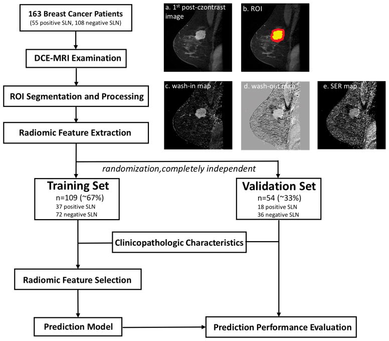
A total of 163 breast cancer patients (55 positive SLN and 108 negative SLN).
FIELD STRENGTH/SEQUENCE: 1.5T, T -weighted DCE-MRI.
A total of 590 radiomic features were extracted for each patient from both intratumoral and peritumoral regions of interest. To avoid overfitting, the dataset was randomly separated into a training set (∼67%) and a validation set (∼33%). The prediction models were built with the training set using logistic regression on the most significant radiomic features in the training set combined with or without clinicopathologic characteristics. The prediction performance was further evaluated in the independent validation set.
Mann-Whitney U-test, Spearman correlation, least absolute shrinkage selection operator (LASSO) regression, logistic regression, and receiver operating characteristic (ROC) analysis were performed.
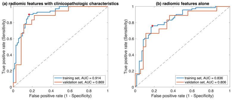
Combining radiomic features with clinicopathologic characteristics, six features were automatically selected in the training set to establish the prediction model of SLN metastasis. In the independent validation set, the area under ROC curve (AUC) was 0.869 (NPV = 0.886). Using radiomic features alone in the same procedure, 4 features were selected and the validation set AUC was 0.806 (NPV = 0.824).
This is the first attempt to demonstrate the feasibility of using DCE-MRI radiomics to predict SLN metastasis in breast cancer. Clinicopathologic characteristics improved the prediction performance. This study provides noninvasive methods to evaluate SLN status for guiding further treatment of breast cancer patients, and can potentially benefit those with negative SLN, by eliminating unnecessary invasive lymph node removal and the associated complications, which is a step further towards precision medicine.
1 Technical Efficacy: Stage 2 J. Magn. Reson. Imaging 2019;49:131-140.
前哨淋巴结(SLN)状态是乳腺癌患者的一个重要预后因素,目前通过有创的 SLN 活检在临床实践中确定。
使用动态对比增强(DCE)磁共振成像(MRI)的肿瘤内和肿瘤周围放射组学特征,结合或不结合原发性肿瘤的临床病理特征,无创预测乳腺癌的 SLN 转移。
回顾性。
共 163 例乳腺癌患者(55 例 SLN 阳性和 108 例 SLN 阴性)。
磁场强度/序列:1.5T,T1 加权 DCE-MRI。
从肿瘤内和肿瘤周围感兴趣区域为每位患者提取了总共 590 个放射组学特征。为了避免过度拟合,数据集随机分为训练集(约 67%)和验证集(约 33%)。使用训练集中最显著的放射组学特征进行逻辑回归,建立预测模型,并结合或不结合临床病理特征,在训练集中进行。在独立的验证集中进一步评估预测性能。
采用 Mann-Whitney U 检验、Spearman 相关分析、最小绝对值收缩选择算子(LASSO)回归、逻辑回归和接收者操作特征(ROC)分析。
将放射组学特征与临床病理特征相结合,在训练集中自动选择了 6 个特征来建立 SLN 转移的预测模型。在独立的验证集中,ROC 曲线下面积(AUC)为 0.869(NPV=0.886)。在相同的步骤中仅使用放射组学特征,选择了 4 个特征,验证集 AUC 为 0.806(NPV=0.824)。
这是首次尝试证明使用 DCE-MRI 放射组学预测乳腺癌 SLN 转移的可行性。临床病理特征提高了预测性能。本研究为评估 SLN 状态提供了非侵入性方法,以指导乳腺癌患者的进一步治疗,并且可以通过消除不必要的有创淋巴结切除和相关并发症,为阴性 SLN 的患者带来潜在益处,这是迈向精准医学的一步。
1 技术功效:2 期 J. Magn. Reson. Imaging 2019;49:131-140.