Institut du Cerveau et de la Moelle épinière, ICM, Sorbonne Université, Inserm, CNRS, AP-HP, F-75013, Paris, France.
Federated Department of Biological Sciences, New Jersey Institute of Technology, University Heights, Newark, NJ, 07102, USA.
Sci Rep. 2018 Sep 11;8(1):13615. doi: 10.1038/s41598-018-31968-4.
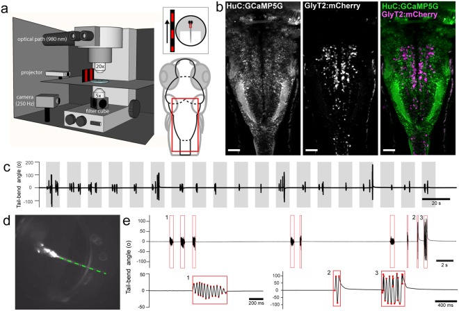
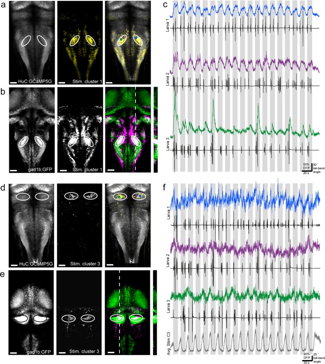
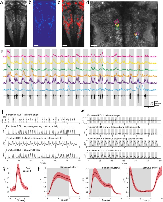
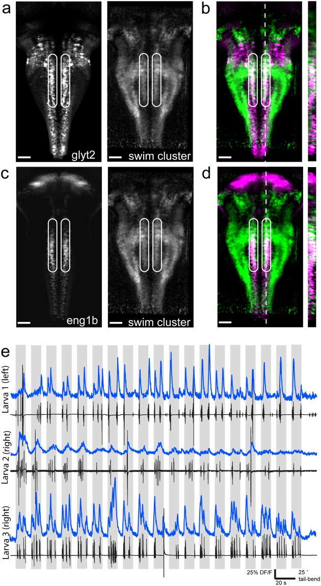
Locomotion in vertebrates relies on motor circuits in the spinal cord receiving inputs from the hindbrain to execute motor commands while dynamically integrating proprioceptive sensory feedback. The spatial organization of the neuronal networks driving locomotion in the hindbrain and role of inhibition has not been extensively investigated. Here, we mapped neuronal activity with single-cell resolution in the hindbrain of restrained transgenic Tg(HuC:GCaMP5G) zebrafish larvae swimming in response to whole-field visual motion. We combined large-scale population calcium imaging in the hindbrain with simultaneous high-speed recording of the moving tail in animals where specific markers label glycinergic inhibitory neurons. We identified cells whose activity preferentially correlates with the visual stimulus or motor activity and used brain registration to compare data across individual larvae. We then morphed calcium imaging data onto the zebrafish brain atlas to compare with known transgenic markers. We report cells localized in the cerebellum whose activity is shut off by the onset of the visual stimulus, suggesting these cells may be constitutively active and silenced during sensorimotor processing. Finally, we discover that the activity of a medial stripe of glycinergic neurons in the domain of expression of the transcription factor engrailed1b is highly correlated with the onset of locomotion. Our efforts provide a high-resolution, open-access dataset for the community by comparing our functional map of the hindbrain to existing open-access atlases and enabling further investigation of this population's role in locomotion.
脊椎动物的运动依赖于脊髓中的运动回路,这些回路接收来自后脑的输入,以执行运动指令,同时动态整合本体感受感觉反馈。驱动后脑运动的神经元网络的空间组织和抑制作用尚未得到广泛研究。在这里,我们在响应全视野视觉运动而游泳的束缚性转基因 Tg(HuC:GCaMP5G)斑马鱼幼虫的后脑中以单细胞分辨率绘制了神经元活动图。我们将后脑中的大规模群体钙成像与动物中特定标记物标记的甘氨酸能抑制性神经元的高速移动尾巴同时记录相结合。我们确定了那些活动与视觉刺激或运动活动优先相关的细胞,并使用大脑注册将数据与个体幼虫进行比较。然后,我们将钙成像数据变形到斑马鱼大脑图谱上,与已知的转基因标记物进行比较。我们报告了定位于小脑的细胞,其活动在视觉刺激开始时被关闭,这表明这些细胞可能在感觉运动处理过程中持续活跃并被抑制。最后,我们发现,在转录因子 engrailed1b 的表达域中,一个中条纹的甘氨酸能神经元的活动与运动的开始高度相关。我们通过将后脑的功能图谱与现有的开放获取图谱进行比较,为社区提供了一个高分辨率的开放获取数据集,并为进一步研究该群体在运动中的作用提供了可能。