• 文献检索
  • 文档翻译
  • 深度研究
  • 学术资讯
  • Suppr Zotero 插件Zotero 插件
  • 邀请有礼
  • 套餐&价格
  • 历史记录
应用&插件
Suppr Zotero 插件Zotero 插件浏览器插件Mac 客户端Windows 客户端微信小程序
定价
高级版会员购买积分包购买API积分包
服务
文献检索文档翻译深度研究API 文档MCP 服务
关于我们
关于 Suppr公司介绍联系我们用户协议隐私条款
关注我们

Suppr 超能文献

核心技术专利:CN118964589B侵权必究
粤ICP备2023148730 号-1Suppr @ 2026

文献检索

告别复杂PubMed语法,用中文像聊天一样搜索,搜遍4000万医学文献。AI智能推荐,让科研检索更轻松。

立即免费搜索

文件翻译

保留排版,准确专业,支持PDF/Word/PPT等文件格式,支持 12+语言互译。

免费翻译文档

深度研究

AI帮你快速写综述,25分钟生成高质量综述,智能提取关键信息,辅助科研写作。

立即免费体验

实时脑肿瘤成像的内源性荧光探针:新鲜人体样本的诊断概念验证研究。

Real-time Brain Tumor imaging with endogenous fluorophores: a diagnosis proof-of-concept study on fresh human samples.

机构信息

IMNC Laboratory, UMR 8165-CNRS/IN2P3, Paris-Saclay university, 91405, Orsay, France.

Neurosurgery Department, Sainte-Anne Hospital, Paris, France.

出版信息

Sci Rep. 2018 Oct 5;8(1):14888. doi: 10.1038/s41598-018-33134-2.

DOI:10.1038/s41598-018-33134-2
PMID:30291269
原文链接:https://pmc.ncbi.nlm.nih.gov/articles/PMC6173695/
Abstract

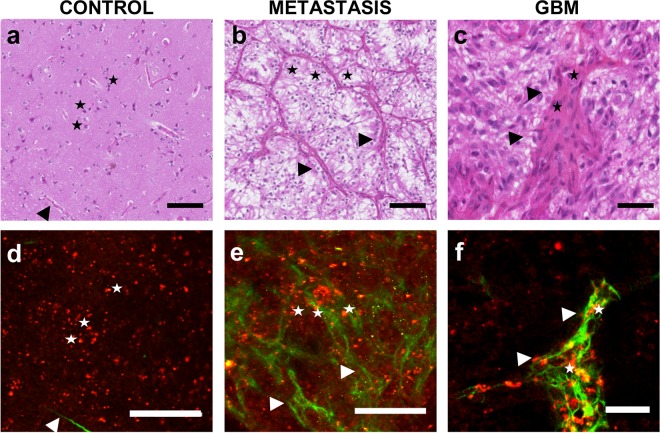
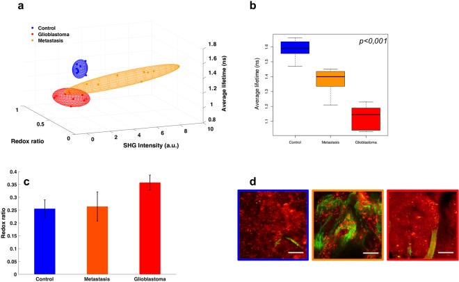
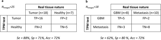
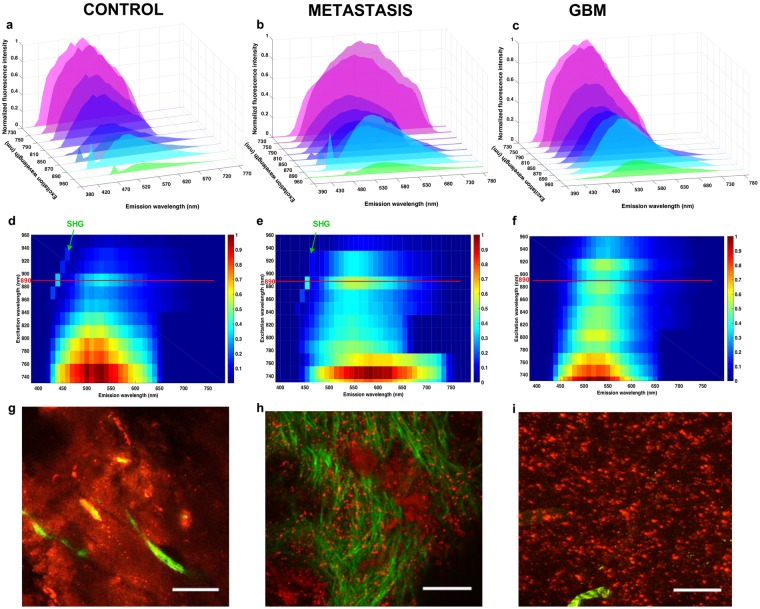
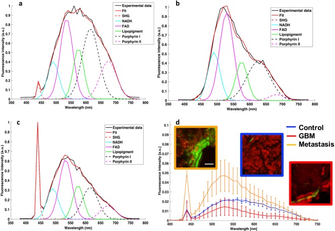
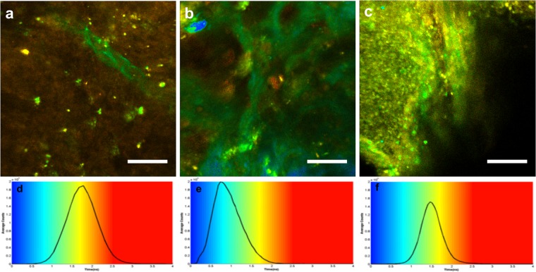
The primary line of therapy for high-grade brain tumor is surgical resection, however, identifying tumor margins in vivo remains a major challenge. Despite the progress in computer-assisted imaging techniques, biopsy analysis remains the standard diagnostic tool when it comes to delineating tumor margins. Our group aims to answer this challenge by exploiting optical imaging of endogenous fluorescence in order to provide a reliable and reproducible diagnosis close to neuropathology. In this study, we first establish the ability of two-photon microscopy (TPM) to discriminate normal brain tissue from glioblastomas and brain metastasis using the endogenous fluorescence response of fresh human brain sample. Two-photon fluorescence images were compared to gold standard neuropathology. "Blind" diagnosis realized by a neuropathologist on a group of TPM images show a good sensitivity, 100%, and specificity, 50% to discriminate non tumoral brain tissue versus glioblastoma or brain metastasis. Quantitative analysis on spectral and fluorescence lifetime measurements resulted in building a scoring system to discriminate brain tissue samples.

摘要

高级别脑肿瘤的主要治疗方法是手术切除,然而,在体内识别肿瘤边界仍然是一个主要挑战。尽管计算机辅助成像技术取得了进展,但活检分析仍然是描绘肿瘤边界的标准诊断工具。我们的研究小组旨在通过利用内源性荧光的光学成像来应对这一挑战,以便提供接近神经病理学的可靠和可重复的诊断。在这项研究中,我们首先使用新鲜人脑样本的内源性荧光反应,建立了双光子显微镜(TPM)区分正常脑组织与胶质母细胞瘤和脑转移的能力。将双光子荧光图像与金标准神经病理学进行比较。神经病理学家对一组 TPM 图像进行的“盲目”诊断显示出良好的敏感性,100%,特异性,50%,可区分非肿瘤性脑组织与胶质母细胞瘤或脑转移。对光谱和荧光寿命测量的定量分析导致建立了一个评分系统来区分脑组织样本。

https://cdn.ncbi.nlm.nih.gov/pmc/blobs/20f9/6173695/403a6536b1a3/41598_2018_33134_Fig6_HTML.jpg
https://cdn.ncbi.nlm.nih.gov/pmc/blobs/20f9/6173695/acd212221394/41598_2018_33134_Fig1_HTML.jpg
https://cdn.ncbi.nlm.nih.gov/pmc/blobs/20f9/6173695/e0c824099b62/41598_2018_33134_Fig2_HTML.jpg
https://cdn.ncbi.nlm.nih.gov/pmc/blobs/20f9/6173695/f291b4b30edc/41598_2018_33134_Fig3_HTML.jpg
https://cdn.ncbi.nlm.nih.gov/pmc/blobs/20f9/6173695/17665fcba4c4/41598_2018_33134_Fig4_HTML.jpg
https://cdn.ncbi.nlm.nih.gov/pmc/blobs/20f9/6173695/137ee5855936/41598_2018_33134_Fig5_HTML.jpg
https://cdn.ncbi.nlm.nih.gov/pmc/blobs/20f9/6173695/403a6536b1a3/41598_2018_33134_Fig6_HTML.jpg

相似文献

1
Real-time Brain Tumor imaging with endogenous fluorophores: a diagnosis proof-of-concept study on fresh human samples.实时脑肿瘤成像的内源性荧光探针:新鲜人体样本的诊断概念验证研究。
Sci Rep. 2018 Oct 5;8(1):14888. doi: 10.1038/s41598-018-33134-2.
2
Multimodal imaging to explore endogenous fluorescence of fresh and fixed human healthy and tumor brain tissues.多模态成像技术探索新鲜和固定的人脑组织中内源性荧光的特性。
J Biophotonics. 2019 Mar;12(3):e201800178. doi: 10.1002/jbio.201800178. Epub 2018 Oct 23.
3
First-in-human intraoperative near-infrared fluorescence imaging of glioblastoma using cetuximab-IRDye800.使用西妥昔单抗-IR800 进行胶质母细胞瘤的首次人体术中近红外荧光成像。
J Neurooncol. 2018 Aug;139(1):135-143. doi: 10.1007/s11060-018-2854-0. Epub 2018 Apr 6.
4
Whole-brain spectroscopic MRI biomarkers identify infiltrating margins in glioblastoma patients.全脑磁共振波谱成像生物标志物可识别胶质母细胞瘤患者的浸润边缘。
Neuro Oncol. 2016 Aug;18(8):1180-9. doi: 10.1093/neuonc/now036. Epub 2016 Mar 15.
5
In vivo multiphoton tomography and fluorescence lifetime imaging of human brain tumor tissue.人脑肿瘤组织的体内多光子断层扫描和荧光寿命成像
J Neurooncol. 2016 May;127(3):473-82. doi: 10.1007/s11060-016-2062-8. Epub 2016 Jan 30.
6
Multiphoton excitation of autofluorescence for microscopy of glioma tissue.用于胶质瘤组织显微镜检查的自发荧光多光子激发
Neurosurgery. 2006 Apr;58(4):759-67; discussion 759-67. doi: 10.1227/01.NEU.0000204885.45644.22.
7
A prospective Phase II clinical trial of 5-aminolevulinic acid to assess the correlation of intraoperative fluorescence intensity and degree of histologic cellularity during resection of high-grade gliomas.一项评估5-氨基乙酰丙酸在高级别胶质瘤切除术中荧光强度与组织学细胞密度相关性的前瞻性II期临床试验。
J Neurosurg. 2016 May;124(5):1300-9. doi: 10.3171/2015.5.JNS1577. Epub 2015 Nov 6.
8
Bioluminescence imaging and two-photon microscopy guided laser ablation of GBM decreases tumor burden.生物发光成像和双光子显微镜引导下的 GBM 激光消融可降低肿瘤负担。
Theranostics. 2018 Jul 16;8(15):4072-4085. doi: 10.7150/thno.25357. eCollection 2018.
9
Optical and nuclear imaging of glioblastoma with phosphatidylserine-targeted nanovesicles.使用磷脂酰丝氨酸靶向纳米囊泡对胶质母细胞瘤进行光学和核成像。
Oncotarget. 2016 May 31;7(22):32866-75. doi: 10.18632/oncotarget.8763.
10
The Impact of Compressed Femtosecond Laser Pulse Durations on Neuronal Tissue Used for Two-Photon Excitation Through an Endoscope.压缩飞秒激光脉冲持续时间对经内窥镜用于双光子激发的神经元组织的影响。
Sci Rep. 2018 Jul 24;8(1):11124. doi: 10.1038/s41598-018-29404-8.

引用本文的文献

1
Towards Optical Biopsy in Glioma Surgery.迈向神经胶质瘤手术中的光学活检
Int J Mol Sci. 2025 May 9;26(10):4554. doi: 10.3390/ijms26104554.
2
Proof of concept study on differentiating metastatic brain samples by their originating organ using multimodal autofluorescence spectroscopy.使用多模态自发荧光光谱法根据转移脑样本的起源器官对其进行区分的概念验证研究。
Sci Rep. 2025 Apr 3;15(1):11476. doi: 10.1038/s41598-025-92366-1.
3
Bi-exponential diffusion-weighted imaging for differentiating high-grade gliomas from solitary brain metastases: a VOI-based histogram analysis.

本文引用的文献

1
Individual Variability of the Human Cerebral Cortex Identified Using Intraoperative Mapping.利用术中映射技术识别人类大脑皮质的个体差异。
World Neurosurg. 2018 Jan;109:e313-e317. doi: 10.1016/j.wneu.2017.09.170. Epub 2017 Oct 5.
2
Neurosurgical management of brain metastases.脑转移瘤的神经外科治疗
Clin Exp Metastasis. 2017 Oct;34(6-7):377-389. doi: 10.1007/s10585-017-9860-z. Epub 2017 Sep 30.
3
Identifying the neck margin status of ductal adenocarcinoma in the pancreatic head by multiphoton microscopy.多光子显微镜鉴别胰头导管腺癌的颈部边界状态。
双指数扩散加权成像用于鉴别高级别胶质瘤与孤立性脑转移瘤:基于感兴趣区的直方图分析
Sci Rep. 2024 Dec 30;14(1):31909. doi: 10.1038/s41598-024-83452-x.
4
Clinical confocal laser endomicroscopy for imaging of autofluorescence signals of human brain tumors and non-tumor brain.用于人脑肿瘤和非肿瘤性脑的自发荧光信号成像的临床共聚焦激光内镜检查术
J Cancer Res Clin Oncol. 2024 Dec 26;151(1):19. doi: 10.1007/s00432-024-06052-2.
5
Multi-Scale Label-Free Human Brain Imaging with Integrated Serial Sectioning Polarization Sensitive Optical Coherence Tomography and Two-Photon Microscopy.基于集成体视学切片偏光敏感光学相干层析成像和双光子显微镜的多尺度无标记人脑成像
Adv Sci (Weinh). 2023 Dec;10(35):e2303381. doi: 10.1002/advs.202303381. Epub 2023 Oct 26.
6
In vivo characterization of the human glioblastoma infiltrative edge with label-free intraoperative fluorescence lifetime imaging.利用无标记术中荧光寿命成像对人胶质母细胞瘤浸润边缘进行体内表征。
Biomed Opt Express. 2023 Apr 24;14(5):2196-2208. doi: 10.1364/BOE.481304. eCollection 2023 May 1.
7
Dysplasia and tumor discrimination in brain tissues by combined fluorescence, Raman, and diffuse reflectance spectroscopies.通过联合荧光、拉曼和漫反射光谱法鉴别脑组织中的发育异常和肿瘤
Biomed Opt Express. 2023 Feb 24;14(3):1256-1275. doi: 10.1364/BOE.477035. eCollection 2023 Mar 1.
8
Comparative Study Between a Customized Bimodal Endoscope and a Benchtop Microscope for Quantitative Tissue Diagnosis.定制双模态内窥镜与台式显微镜用于定量组织诊断的比较研究
Front Oncol. 2022 May 12;12:881331. doi: 10.3389/fonc.2022.881331. eCollection 2022.
9
Creation of Non-Contact Device for Use in Metastatic Melanoma Margin Identification in Mouse Brain.用于小鼠脑转移黑色素瘤边缘识别的非接触式设备的创建。
Proc SPIE Int Soc Opt Eng. 2022 Jan-Feb;11945. doi: 10.1117/12.2608975. Epub 2022 Mar 4.
10
Advances in nonlinear optical microscopy techniques for in vivo and in vitro neuroimaging.用于体内和体外神经成像的非线性光学显微镜技术进展。
Biophys Rev. 2021 Aug 31;13(6):1199-1217. doi: 10.1007/s12551-021-00832-7. eCollection 2021 Dec.
Sci Rep. 2017 Jul 4;7(1):4586. doi: 10.1038/s41598-017-04771-w.
4
Direct electrical bipolar electrostimulation for functional cortical and subcortical cerebral mapping in awake craniotomy. Practical considerations.清醒开颅手术中用于功能性皮质和皮质下脑图谱绘制的直接双极电刺激。实际考量
Neurochirurgie. 2017 Jun;63(3):164-174. doi: 10.1016/j.neuchi.2016.08.009. Epub 2017 Feb 2.
5
Fluorescence-guided resection of brain tumor: review of the significance of intraoperative quantification of protoporphyrin IX fluorescence.荧光引导下脑肿瘤切除术:术中原卟啉IX荧光定量的意义综述
Neurophotonics. 2017 Jan;4(1):011011. doi: 10.1117/1.NPh.4.1.011011. Epub 2017 Jan 12.
6
Spectral and fluorescence lifetime endoscopic system using a double-clad photonic crystal fiber.使用双包层光子晶体光纤的光谱和荧光寿命内镜系统。
Opt Lett. 2016 Nov 15;41(22):5214-5217. doi: 10.1364/OL.41.005214.
7
Optical redox ratio and endogenous porphyrins in the detection of urinary bladder cancer: A patient biopsy analysis.光学氧化还原比率和内源性卟啉在膀胱癌检测中的应用:患者活检分析
J Biophotonics. 2017 Aug;10(8):1062-1073. doi: 10.1002/jbio.201600162. Epub 2016 Oct 7.
8
Characterization of fiber ultrashort pulse delivery for nonlinear endomicroscopy.用于非线性内镜显微镜检查的光纤超短脉冲传输特性
Opt Express. 2016 Jun 13;24(12):12515-23. doi: 10.1364/OE.24.012515.
9
Maximizing safe resection of low- and high-grade glioma.最大限度安全切除低级别和高级别胶质瘤。
J Neurooncol. 2016 Nov;130(2):269-282. doi: 10.1007/s11060-016-2110-4. Epub 2016 May 12.
10
Two-photon fluorescence lifetime imaging of primed SNARE complexes in presynaptic terminals and β cells.突触前终末和β细胞中引发的SNARE复合体的双光子荧光寿命成像。
Nat Commun. 2015 Oct 6;6:8531. doi: 10.1038/ncomms9531.