Brigham and Women's Hospital, Harvard Medical School, USA.
Massachusetts General Hospital, Harvard Medical School, USA.
Brain Stimul. 2019 Jan-Feb;12(1):129-138. doi: 10.1016/j.brs.2018.10.004. Epub 2018 Oct 13.
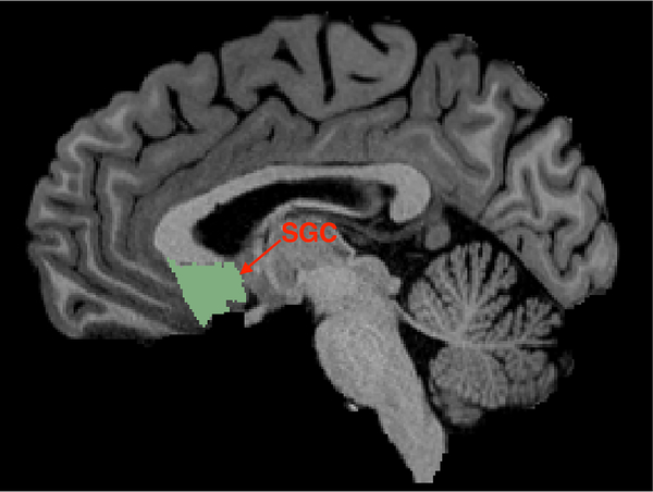
Transcranial magnetic stimulation (TMS) is a noninvasive neuromodulation technique with therapeutic applications for the treatment of major depressive disorder (MDD). The standard protocol uses high frequency stimulation over the left dorsolateral prefrontal cortex (DLPFC) identified in a heuristic manner leading to moderate clinical efficacy. A proposed strategy to increase the anatomical precision in targeting, based on resting-state functional MRI (rsfMRI), identifies the subregion within the DLPFC having the strongest anticorrelated functional connectivity with the subgenual cortex (SGC) for each individual subject.
In this work, we comprehensively test the reliability and reproducibility of this targeting method for different scan lengths on 100 subjects from the Human Connectome Project (HCP) where each subject had a four 15-min rsfMRI scan on 2 different days.
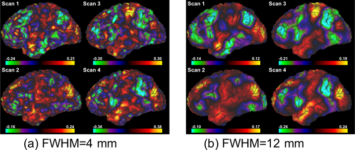
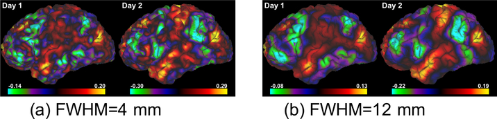
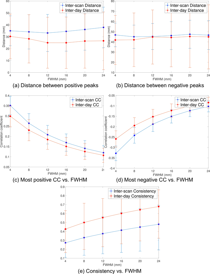
We quantified the inter-scan and inter-day distance between the rsfMRI-guided DLPFC targets for each subject controlling for a number of expected sources of noise using volumetric as well as surface analyses.
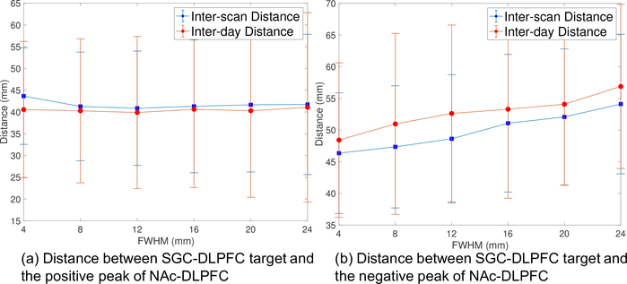
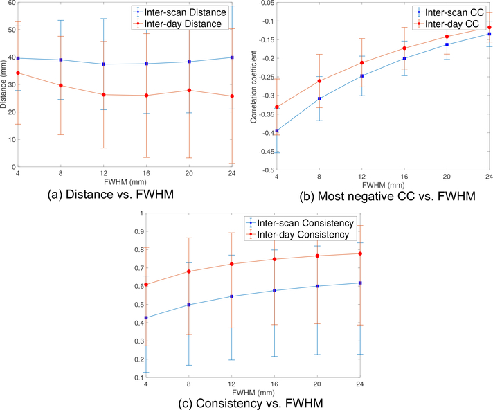
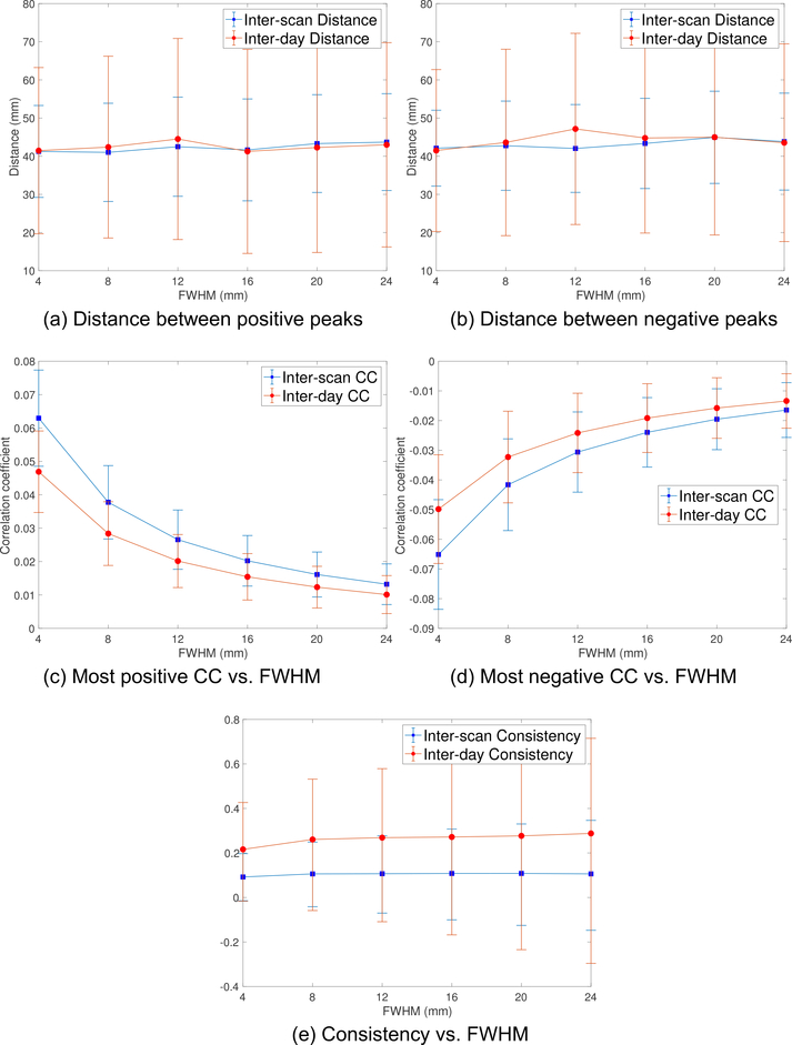
Our results show that the average inter-day distance (with fMRI scans lasting 30 min on each day) is 25% less variable than the inter-scan distance, which uses 50% less data. Specifically, the inter-scan distance was more than 37 mm, while for the longer-scan, the inter-day distance had lower variability at 25 mm. Finally, we tested the same rsfMRI strategy using the nucleus accumbens (NAc) as a control region relevant to MDD but less susceptible to artifacts, using both volume and surface rsfMRI data. The results showed similar variability to the SGC-DLPFC functional connectivity. Moreover, our results suggest that a smoothing kernel with 12 mm full-width half maximum (FWHM) lead to more stable and reliable target estimates.
Our work provides a quantitative assessment of the topographic precision of this targeting method, describing an anatomical variability that may surpass the spatial resolution of some forms of focal TMS as it is commonly applied, and provides recommendations for improved accuracy.
经颅磁刺激(TMS)是一种非侵入性神经调节技术,具有治疗重度抑郁症(MDD)的应用。标准方案使用启发式方法在左侧背外侧前额叶皮层(DLPFC)上进行高频刺激,从而产生中等的临床疗效。一种基于静息态功能磁共振成像(rsfMRI)的增加靶向解剖精度的建议策略,为每个个体识别 DLPFC 内与 subgenual 皮层(SGC)具有最强反相关功能连接的子区域。
在这项工作中,我们使用来自人类连接组计划(HCP)的 100 名受试者的 100 名受试者,对不同扫描长度的这种靶向方法的可靠性和可重复性进行了全面测试,其中每个受试者在 2 天的 4 次 15 分钟 rsfMRI 扫描。
我们使用容积和表面分析控制许多预期的噪声源,对每个受试者的 rsfMRI 引导的 DLPFC 目标的扫描间和扫描内距离进行量化。
我们的结果表明,与使用 50%的数据相比,平均扫描内距离(每天 fMRI 扫描持续 30 分钟)的变异性降低了 25%。具体来说,扫描间距离超过 37mm,而对于较长的扫描,扫描内距离的变异性在 25mm 时较低。最后,我们使用与 MDD 相关但较少受伪影影响的核壳(NAc)作为对照区域,测试了相同的 rsfMRI 策略,使用了容积和表面 rsfMRI 数据。结果显示与 SGC-DLPFC 功能连接相似的可变性。此外,我们的结果表明,使用 12mm 全宽半最大值(FWHM)的平滑核可导致更稳定和可靠的目标估计。
我们的工作提供了对这种靶向方法的拓扑精度的定量评估,描述了一种解剖学变异性,这种变异性可能超过某些形式的聚焦 TMS 的空间分辨率,因为它通常应用,并提供了提高准确性的建议。