文献检索文档翻译深度研究
Suppr Zotero 插件Zotero 插件
邀请有礼套餐&价格历史记录

新学期,新优惠

限时优惠:9月1日-9月22日

30天高级会员仅需29元

1天体验卡首发特惠仅需5.99元

了解详情
不再提醒
插件&应用
Suppr Zotero 插件Zotero 插件浏览器插件Mac 客户端Windows 客户端微信小程序
高级版
套餐订阅购买积分包
AI 工具
文献检索文档翻译深度研究
关于我们
关于 Suppr公司介绍联系我们用户协议隐私条款
关注我们

Suppr 超能文献

核心技术专利:CN118964589B侵权必究
粤ICP备2023148730 号-1Suppr @ 2025

基于 MRI 的宫颈癌放射组学列线图术前预测淋巴结脉管间隙侵犯

MR-Based Radiomics Nomogram of Cervical Cancer in Prediction of the Lymph-Vascular Space Invasion preoperatively.

机构信息

Department of Radiology, Shanghai General Hospital, Shanghai Jiao Tong University School of Medicine, Shanghai, P.R. China.

CAS Key Laboratory of Molecular Imaging, Institute of Automation, Chinese Academy of Sciences, Beijing, P.R. China.

出版信息

J Magn Reson Imaging. 2019 May;49(5):1420-1426. doi: 10.1002/jmri.26531. Epub 2018 Oct 26.


DOI:10.1002/jmri.26531
PMID:30362652
原文链接:https://pmc.ncbi.nlm.nih.gov/articles/PMC6587470/
Abstract

BACKGROUND: Lymph-vascular space invasion (LVSI) is an unfavorable prognostic factor in cervical cancer. Unfortunately, there are no current clinical tools for the preoperative prediction of LVSI. PURPOSE: 
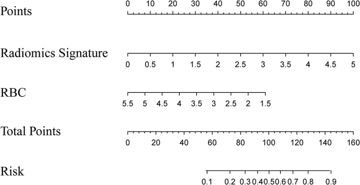
To develop and validate an axial T contrast-enhanced (CE) MR-based radiomics nomogram that incorporated a radiomics signature and some clinical parameters for predicting LVSI of cervical cancer preoperatively. STUDY TYPE: Retrospective. POPULATION: In all, 105 patients were randomly divided into two cohorts at a 2:1 ratio. FIELD STRENGTH/SEQUENCE: T CE MRI sequences at 1.5T. ASSESSMENT: 
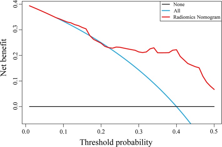
Univariate analysis was performed on the radiomics features and clinical parameters. Multivariate analysis was performed to determine the optimal feature subset. The receiver operating characteristic (ROC) analysis was performed to evaluate the performance of prediction model and radiomics nomogram. STATISTICAL TESTS: 
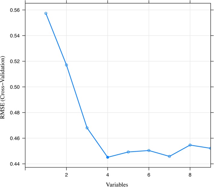
The Mann-Whitney U-test and the chi-square test were used to evaluate the performance of clinical characteristics and LVSI status by pathology. The minimum-redundancy/maximum-relevance and recursive feature elimination methods were applied to select the features. The radiomics model was constructed using logistic regression. RESULTS: 
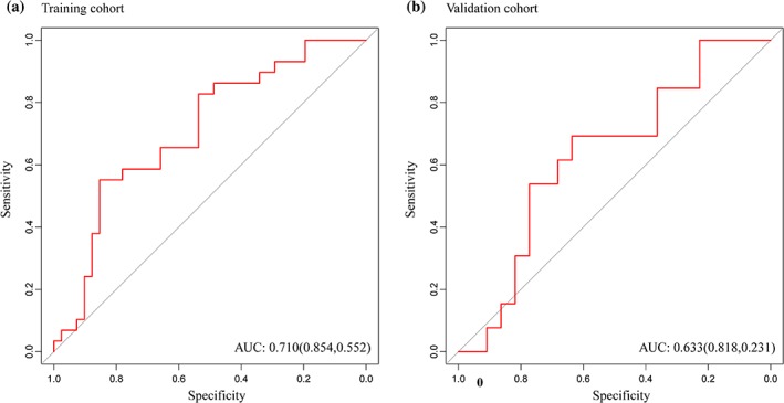
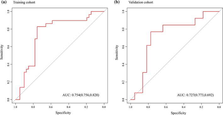
Three radiomics features and one clinical characteristic were selected. The radiomics nomogram showed favorable discrimination between LVSI and non-LVSI groups. The AUC was 0.754 (95% confidence interval [CI], 0.6326-0.8745) in the training cohort and 0.727 (95% CI, 0.5449-0.9097) in the validation cohort. The specificity and sensitivity were 0.756 and 0.828 in the training cohort and 0.773 and 0.692 in the validation cohort. DATA CONCLUSION: T CE MR-based radiomics nomogram serves as a noninvasive biomarker in the prediction of LVSI in patients with cervical cancer preoperatively. LEVEL OF EVIDENCE: 4 Technical Efficacy: Stage 2 J. Magn. Reson. Imaging 2019;49:1420-1426.

摘要

背景:淋巴血管空间侵犯(LVSI)是宫颈癌的一个不利预后因素。不幸的是,目前还没有用于术前预测 LVSI 的临床工具。

目的:开发和验证一种基于轴位 T 对比增强(CE)MR 的放射组学列线图,该列线图结合了放射组学特征和一些临床参数,用于术前预测宫颈癌的 LVSI。

研究类型:回顾性。

人群:总共,105 名患者以 2:1 的比例随机分为两组。

磁场强度/序列:1.5T 的 T CE MRI 序列。

评估:对放射组学特征和临床参数进行单变量分析。进行多变量分析以确定最佳特征子集。通过接收者操作特征(ROC)分析评估预测模型和放射组学列线图的性能。

统计学检验:Mann-Whitney U 检验和卡方检验用于评估临床特征和病理 LVSI 状态的性能。应用最小冗余/最大相关性和递归特征消除方法来选择特征。使用逻辑回归构建放射组学模型。

结果:选择了三个放射组学特征和一个临床特征。放射组学列线图显示出区分 LVSI 和非 LVSI 组的良好能力。在训练队列中的 AUC 为 0.754(95%置信区间[CI],0.6326-0.8745),在验证队列中的 AUC 为 0.727(95%CI,0.5449-0.9097)。在训练队列中的特异性和敏感性分别为 0.756 和 0.828,在验证队列中的特异性和敏感性分别为 0.773 和 0.692。

数据结论:基于 T CE MR 的放射组学列线图可作为预测宫颈癌患者术前 LVSI 的非侵入性生物标志物。

证据水平:4 技术功效:第 2 阶段 J. Magn. Reson. Imaging 2019;49:1420-1426。

https://cdn.ncbi.nlm.nih.gov/pmc/blobs/1402/6587470/c33be4ce6ffe/JMRI-49-1420-g005.jpg
https://cdn.ncbi.nlm.nih.gov/pmc/blobs/1402/6587470/ae38b2596f67/JMRI-49-1420-g001.jpg
https://cdn.ncbi.nlm.nih.gov/pmc/blobs/1402/6587470/eafa4fad49bb/JMRI-49-1420-g002.jpg
https://cdn.ncbi.nlm.nih.gov/pmc/blobs/1402/6587470/d943933ecb11/JMRI-49-1420-g003.jpg
https://cdn.ncbi.nlm.nih.gov/pmc/blobs/1402/6587470/f4aff6ee2f30/JMRI-49-1420-g004.jpg
https://cdn.ncbi.nlm.nih.gov/pmc/blobs/1402/6587470/c33be4ce6ffe/JMRI-49-1420-g005.jpg

相似文献

[1]
MR-Based Radiomics Nomogram of Cervical Cancer in Prediction of the Lymph-Vascular Space Invasion preoperatively.

J Magn Reson Imaging. 2018-10-26

[2]
A Multicenter Study on Preoperative Assessment of Lymphovascular Space Invasion in Early-Stage Cervical Cancer Based on Multimodal MR Radiomics.

J Magn Reson Imaging. 2023-11

[3]
Multiparametric MRI-Based Radiomics Nomogram for Predicting Lymph Node Metastasis in Early-Stage Cervical Cancer.

J Magn Reson Imaging. 2020-9

[4]
Multi-parametric MRI-based peritumoral radiomics on prediction of lymph-vascular space invasion in early-stage cervical cancer.

Diagn Interv Radiol. 2022-7

[5]
Multiparametric MRI-Based Radiomics Nomogram for Predicting Lymphovascular Space Invasion in Endometrial Carcinoma.

J Magn Reson Imaging. 2020-10

[6]
Radiomic signature as a predictive factor for lymph node metastasis in early-stage cervical cancer.

J Magn Reson Imaging. 2018-8-13

[7]
Preoperative prediction of lymphovascular invasion in invasive breast cancer with dynamic contrast-enhanced-MRI-based radiomics.

J Magn Reson Imaging. 2019-2-17

[8]
Multiparametric mri-based radiomics nomogram for predicting lymph-vascular space invasion in cervical cancer.

BMC Med Imaging. 2024-7-5

[9]
Multiparametric MRI radiomics nomogram for predicting lymph-vascular space invasion in early-stage cervical cancer.

Br J Radiol. 2022-6-1

[10]
Preoperative prediction of pelvic lymph nodes metastasis in early-stage cervical cancer using radiomics nomogram developed based on T2-weighted MRI and diffusion-weighted imaging.

Eur J Radiol. 2019-3-20

引用本文的文献

[1]
Multi-parametric MRI-based radiomics nomogram for predicting lymphovascular space invasion in early-stage cervical adenocarcinoma.

Front Oncol. 2025-8-21

[2]
Prediction of cervical cancer lymph node metastasis based on multisequence magnetic resonance imaging radiomics and deep learning features: a dual-center study.

Sci Rep. 2025-8-10

[3]
Predicting lymphovascular space invasion in early-stage cervical squamous cell carcinoma using heart rate variability.

Front Oncol. 2025-7-21

[4]
Imaging-Based AI for Predicting Lymphovascular Space Invasion in Cervical Cancer: Systematic Review and Meta-Analysis.

J Med Internet Res. 2025-6-16

[5]
Clinical-radiomics nomogram construction from magnetic resonance imaging to diagnose osteoporosis: a preliminary study.

Eur Spine J. 2025-5-29

[6]
Nomogram prediction of the lymph-vascular space invasion in cervical cancer: comparison of 2009 and 2018 staging systems.

Front Oncol. 2025-3-6

[7]
Deep transfer learning radiomics for distinguishing sinonasal malignancies: a preliminary MRI study.

Future Oncol. 2025-4

[8]
Radiomics based on MRI in predicting lymphovascular space invasion of cervical cancer: a meta-analysis.

Front Oncol. 2024-10-17

[9]
Ranking attention multiple instance learning for lymph node metastasis prediction on multicenter cervical cancer MRI.

J Appl Clin Med Phys. 2024-12

[10]
The role of radiomics for predicting of lymph-vascular space invasion in cervical cancer patients based on artificial intelligence: a systematic review and meta-analysis.

J Gynecol Oncol. 2025-3

本文引用的文献

[1]
Computational Radiomics System to Decode the Radiographic Phenotype.

Cancer Res. 2017-11-1

[2]
Transvaginal ultrasound versus magnetic resonance imaging for preoperative assessment of myometrial infiltration in patients with endometrial cancer: a systematic review and meta-analysis.

J Gynecol Oncol. 2017-11

[3]
Somatic Mutations Drive Distinct Imaging Phenotypes in Lung Cancer.

Cancer Res. 2017-7-15

[4]
MRI features predict survival and molecular markers in diffuse lower-grade gliomas.

Neuro Oncol. 2017-6-1

[5]
Radiomics Features of Multiparametric MRI as Novel Prognostic Factors in Advanced Nasopharyngeal Carcinoma.

Clin Cancer Res. 2017-3-9

[6]
Association between tumor heterogeneity and progression-free survival in non-small cell lung cancer patients with EGFR mutations undergoing tyrosine kinase inhibitors therapy.

Annu Int Conf IEEE Eng Med Biol Soc. 2016-8

[7]
CT-based radiomics signature: a potential biomarker for preoperative prediction of early recurrence in hepatocellular carcinoma.

Abdom Radiol (NY). 2017-6

[8]
Associations Between Somatic Mutations and Metabolic Imaging Phenotypes in Non-Small Cell Lung Cancer.

J Nucl Med. 2017-4

[9]
Radiomics Signature: A Potential Biomarker for the Prediction of Disease-Free Survival in Early-Stage (I or II) Non-Small Cell Lung Cancer.

Radiology. 2016-6-27

[10]
Development and Validation of a Radiomics Nomogram for Preoperative Prediction of Lymph Node Metastasis in Colorectal Cancer.

J Clin Oncol. 2016-5-2

文献AI研究员

20分钟写一篇综述,助力文献阅读效率提升50倍

立即体验

用中文搜PubMed

大模型驱动的PubMed中文搜索引擎

马上搜索

推荐工具

医学文档翻译智能文献检索