Qu Yueqiao, He Youmin, Zhang Yi, Ma Teng, Zhu Jiang, Miao Yusi, Dai Cuixia, Humayun Mark, Zhou Qifa, Chen Zhongping
Beckman Laser Institute, University of California, Irvine, 1002 Health Sciences Road East, Irvine, CA 92612, USA.
First two authors contributed equally to this work.
Biomed Opt Express. 2018 Aug 2;9(9):4054-4063. doi: 10.1364/BOE.9.004054. eCollection 2018 Sep 1.
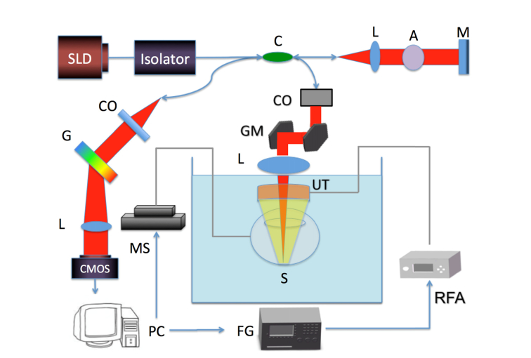
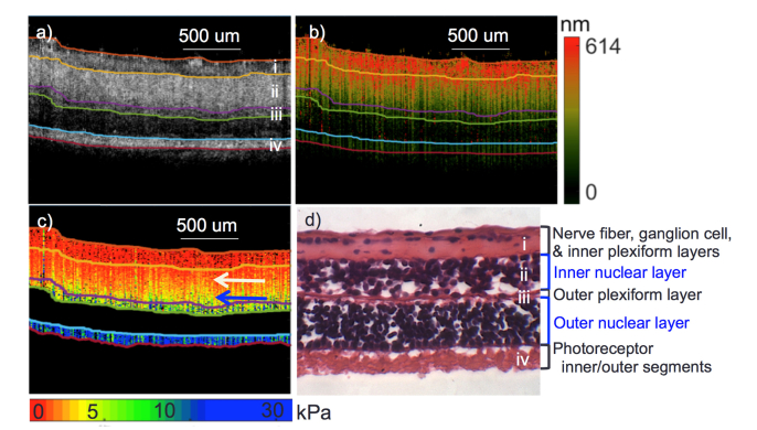
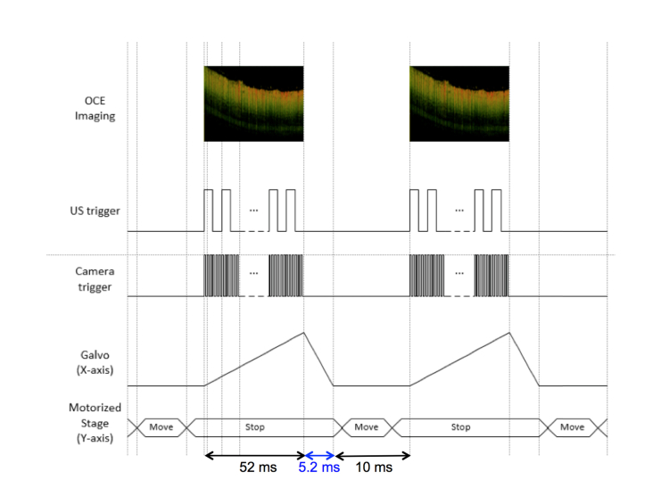
Age-related macular degeneration (AMD) is the leading cause of blindness in the elderly (over the age of 60 years) in western countries. In the early stages of the disease, structural changes may be subtle and cannot be detected. Recently it has been postulated that the mechanical properties of the retina may change with the onset of AMD. In this manuscript, we present a novel, non-invasive means that utilizes synchronized acoustic radiation force optical coherence elastography (ARF-OCE) to measure and estimate the elasticity of cadaver porcine retina. Both regions near the optic nerve and in the peripheral retina were studied. An acoustic force is exerted on the tissue for excitation and the resulting tissue vibrations, often in the nanometer scale, are detected with high-resolution optical methods. Segmentation has been performed to isolate individual layers and the Young's modulus has been estimated for each. The results have been successfully compared and mapped to corresponding histological results using H&E staining. Finally, 64 elastograms of the retina were analyzed, as well as the elastic properties, with stiffness ranging from 1.3 to 25.9 kPa in the ganglion to the photoreceptor sides respectively. ARF-OCE allows for the elasticity mapping of anatomical retinal layers. This imaging approach needs further evaluation but has the potential to allow physicians to gain a better understanding of the elasticity of retinal layers in retinal diseases such as AMD.
年龄相关性黄斑变性(AMD)是西方国家60岁以上老年人失明的主要原因。在疾病的早期阶段,结构变化可能很细微,难以检测到。最近有人推测,随着AMD的发生,视网膜的力学性能可能会发生变化。在本论文中,我们提出了一种新颖的非侵入性方法,该方法利用同步声辐射力光学相干弹性成像(ARF-OCE)来测量和估计猪尸体视网膜的弹性。我们研究了视神经附近和周边视网膜区域。对组织施加声力以进行激发,并使用高分辨率光学方法检测通常在纳米尺度上产生的组织振动。进行了分割以分离各个层,并估计了每层的杨氏模量。使用苏木精和伊红(H&E)染色,已成功将结果与相应的组织学结果进行了比较和映射。最后,分析了64张视网膜弹性成像图以及弹性特性,从神经节侧到光感受器侧的刚度范围分别为1.3至25.9kPa。ARF-OCE可实现视网膜各层的弹性成像。这种成像方法需要进一步评估,但有可能让医生更好地了解诸如AMD等视网膜疾病中视网膜各层的弹性。