He Youmin, Qu Yueqiao, Zhu Jiang, Zhang Yi, Saidi Arya, Ma Teng, Zhou Qifa, Chen Zhongping
Department of Biomedical Engineering and the Beckman Laser Institute, University of California, Irvine, CA 92697 USA.
Roski Eye Institute, Department of Ophthalmology and Biomedical Engineering, University of Southern California, Los Angeles, CA 90089 USA.
IEEE J Sel Top Quantum Electron. 2019 Jan-Feb;25(1). doi: 10.1109/jstqe.2018.2834435. Epub 2018 May 8.
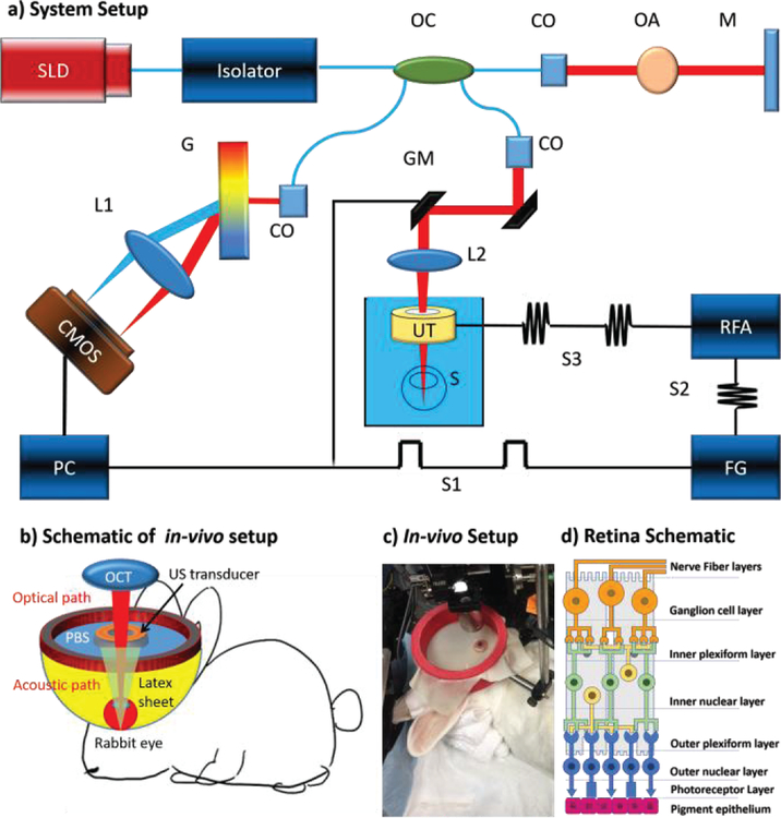
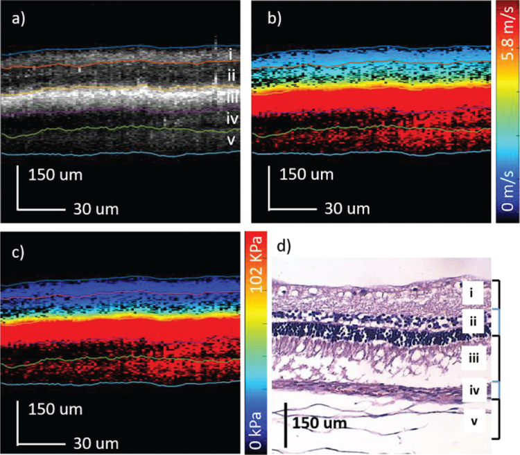
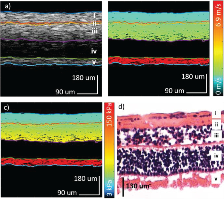
Retinal diseases, such as age-related macular degeneration (AMD), are the leading cause of blindness in the elderly population. Since no known cures are currently present, it is crucial to diagnose the condition in its early stages so that disease progression is monitored. Recent advances show that the mechanical elasticity of the posterior eye changes with the onset of AMD. In this work, we present a quantitative method of mapping the mechanical elasticity of the posterior eye using confocal shear wave acoustic radiation force optical coherence elastography (SW-ARF-OCE). This technique has been developed and validated with both an ex-vivo porcine tissue model and a customized in-vivo rabbit model, which both showed the quantified elasticity variations between different layers. This study verifies the feasibility of using this technology for the quantification and diagnosis of retinal diseases from the in-vivo posterior eye.
视网膜疾病,如年龄相关性黄斑变性(AMD),是老年人群失明的主要原因。由于目前尚无已知的治愈方法,在疾病早期进行诊断以便监测疾病进展至关重要。最近的研究进展表明,随着AMD的发生,眼球后部的机械弹性会发生变化。在这项工作中,我们提出了一种使用共聚焦剪切波声辐射力光学相干弹性成像(SW-ARF-OCE)来绘制眼球后部机械弹性的定量方法。该技术已在离体猪组织模型和定制的活体兔模型上得到开发和验证,这两种模型均显示了不同层之间的弹性变化量化情况。本研究验证了使用该技术对活体眼球后部视网膜疾病进行量化和诊断的可行性。