• 文献检索
  • 文档翻译
  • 深度研究
  • 学术资讯
  • Suppr Zotero 插件Zotero 插件
  • 邀请有礼
  • 套餐&价格
  • 历史记录
应用&插件
Suppr Zotero 插件Zotero 插件浏览器插件Mac 客户端Windows 客户端微信小程序
定价
高级版会员购买积分包购买API积分包
服务
文献检索文档翻译深度研究API 文档MCP 服务
关于我们
关于 Suppr公司介绍联系我们用户协议隐私条款
关注我们

Suppr 超能文献

核心技术专利:CN118964589B侵权必究
粤ICP备2023148730 号-1Suppr @ 2026

文献检索

告别复杂PubMed语法,用中文像聊天一样搜索,搜遍4000万医学文献。AI智能推荐,让科研检索更轻松。

立即免费搜索

文件翻译

保留排版,准确专业,支持PDF/Word/PPT等文件格式,支持 12+语言互译。

免费翻译文档

深度研究

AI帮你快速写综述,25分钟生成高质量综述,智能提取关键信息,辅助科研写作。

立即免费体验

利用光学相干断层扫描血管造影术比较白点综合征继发脉络膜新生血管与年龄相关性黄斑变性

Comparison of choroidal neovascularization secondary to white dot syndromes and age-related macular degeneration by using optical coherence tomography angiography.

作者信息

Wang Jay C, McKay Kenneth M, Sood Arjun B, Laíns Inês, Sobrin Lucia, Miller John B

机构信息

Retina Service, Department of Ophthalmology, Massachusetts Eye and Ear, Harvard Medical School, Boston, MA 02114, USA,

出版信息

Clin Ophthalmol. 2018 Dec 31;13:95-105. doi: 10.2147/OPTH.S185468. eCollection 2019.

DOI:10.2147/OPTH.S185468
PMID:30643383
原文链接:https://pmc.ncbi.nlm.nih.gov/articles/PMC6318713/
Abstract

PURPOSE

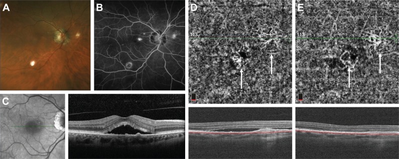
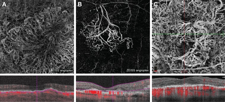
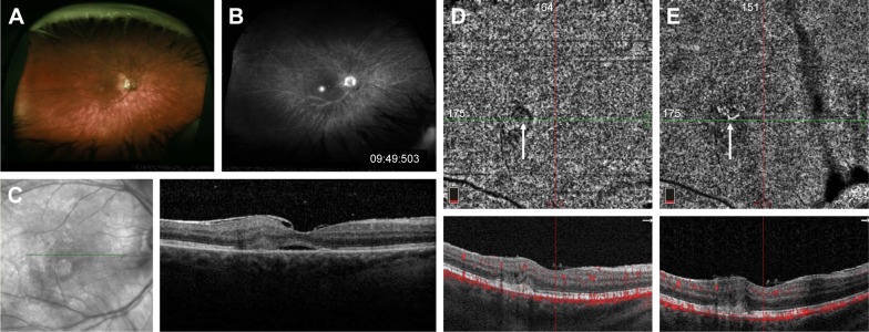
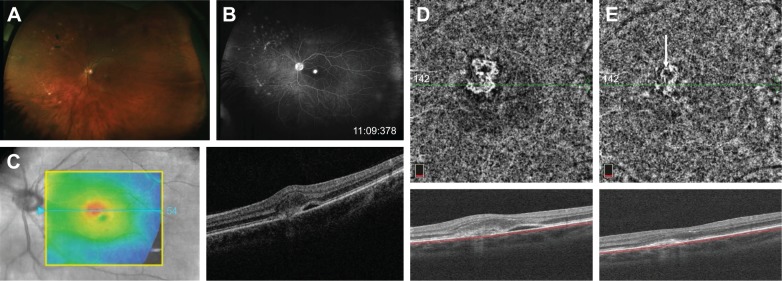
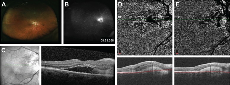
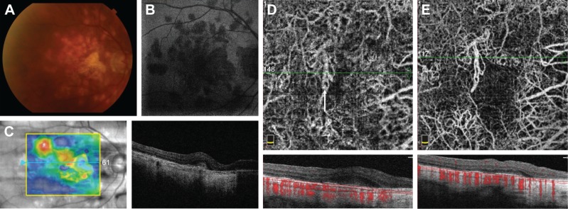
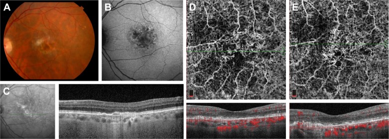
To characterize and compare choroidal neovascularization (CNV) secondary to white dot syndromes (WDS) and age-related macular degeneration (AMD) using optical coherence tomography angiography (OCT-A).

METHODS

This is a cross-sectional study in which we imaged patients with CNV secondary to WDS and AMD with either the Zeiss Angioplex OCT-A or Optovue AngioVue OCT-A. Relevant demographic and clinical characteristics were collected and analyzed. CNV area and vessel density (VD) were measured by three independent graders, and linear regression analysis was subsequently performed.

RESULTS

Three patients with multifocal choroiditis and panuveitis, one patient each with birdshot chorioretinopathy, presumed ocular histoplasmosis syndrome, and persistent placoid maculopathy, and eleven patients with AMD with sufficient image quality were included. CNV associated with WDS was significantly smaller than that secondary to AMD (0.56±0.32 vs 2.79±1.80 mm, =-2.22, =0.01), while no difference in VD was detected (0.46±0.09 vs 0.44±0.09, =0.02, =0.71).

CONCLUSION

CNV networks secondary to WDS appear to be smaller than those secondary to AMD but have similar VD. OCT-A is a powerful tool to investigate properties of CNV from various etiologies. Larger studies are needed for further characterization and understanding of CNV pathogenesis in inflammatory conditions.

摘要

目的

使用光学相干断层扫描血管造影(OCT-A)对白点综合征(WDS)和年龄相关性黄斑变性(AMD)继发的脉络膜新生血管(CNV)进行特征描述和比较。

方法

这是一项横断面研究,我们使用蔡司Angioplex OCT-A或Optovue AngioVue OCT-A对WDS和AMD继发CNV的患者进行成像。收集并分析相关的人口统计学和临床特征。由三名独立的分级人员测量CNV面积和血管密度(VD),随后进行线性回归分析。

结果

纳入了3例多灶性脉络膜炎和全葡萄膜炎患者、1例鸟枪弹样脉络膜视网膜病变患者、1例疑似眼组织胞浆菌病综合征患者、1例持续性地图状黄斑病变患者以及11例图像质量足够的AMD患者。与WDS相关的CNV明显小于AMD继发的CNV(0.56±0.32 vs 2.79±1.80 mm,=-2.22,=0.01),而VD未检测到差异(0.46±0.09 vs 0.44±0.09,=0.02,=0.71)。

结论

WDS继发的CNV网络似乎比AMD继发的小,但VD相似。OCT-A是研究各种病因引起的CNV特性的有力工具。需要进行更大规模的研究以进一步表征和理解炎症性疾病中CNV的发病机制。

https://cdn.ncbi.nlm.nih.gov/pmc/blobs/b53f/6318713/a7418f0ce965/opth-13-095Fig7.jpg
https://cdn.ncbi.nlm.nih.gov/pmc/blobs/b53f/6318713/2a5d88a88acb/opth-13-095Fig1.jpg
https://cdn.ncbi.nlm.nih.gov/pmc/blobs/b53f/6318713/3227de7c83d0/opth-13-095Fig2.jpg
https://cdn.ncbi.nlm.nih.gov/pmc/blobs/b53f/6318713/c998b17dd8e2/opth-13-095Fig3.jpg
https://cdn.ncbi.nlm.nih.gov/pmc/blobs/b53f/6318713/9bb68bfd4a9a/opth-13-095Fig4.jpg
https://cdn.ncbi.nlm.nih.gov/pmc/blobs/b53f/6318713/8963eb8b9207/opth-13-095Fig5.jpg
https://cdn.ncbi.nlm.nih.gov/pmc/blobs/b53f/6318713/d419d0cf3c87/opth-13-095Fig6.jpg
https://cdn.ncbi.nlm.nih.gov/pmc/blobs/b53f/6318713/a7418f0ce965/opth-13-095Fig7.jpg

相似文献

1
Comparison of choroidal neovascularization secondary to white dot syndromes and age-related macular degeneration by using optical coherence tomography angiography.利用光学相干断层扫描血管造影术比较白点综合征继发脉络膜新生血管与年龄相关性黄斑变性
Clin Ophthalmol. 2018 Dec 31;13:95-105. doi: 10.2147/OPTH.S185468. eCollection 2019.
2
Quantitative optical coherence tomography angiography of choroidal neovascularization in age-related macular degeneration.年龄相关性黄斑变性脉络膜新生血管的定量光相干断层扫描血管造影。
Ophthalmology. 2014 Jul;121(7):1435-44. doi: 10.1016/j.ophtha.2014.01.034. Epub 2014 Mar 27.
3
Distinguishing White Dot Syndromes With Patterns of Choroidal Hypoperfusion on Optical Coherence Tomography Angiography.通过光学相干断层扫描血管造影术上脉络膜低灌注模式鉴别白点综合征
Ophthalmic Surg Lasers Imaging Retina. 2017 Aug 1;48(8):638-646. doi: 10.3928/23258160-20170802-06.
4
CHOROIDAL NEOVASCULAR AREA AND VESSEL DENSITY COMPARISON BETWEEN TWO SWEPT-SOURCE OPTICAL COHERENCE TOMOGRAPHY ANGIOGRAPHY DEVICES.两种扫频源光学相干断层血管成像设备的脉络膜新生血管区和血管密度比较。
Retina. 2020 Mar;40(3):521-528. doi: 10.1097/IAE.0000000000002430.
5
CHARACTERIZING THE EFFECT OF ANTI-VASCULAR ENDOTHELIAL GROWTH FACTOR THERAPY ON TREATMENT-NAIVE CHOROIDAL NEOVASCULARIZATION USING OPTICAL COHERENCE TOMOGRAPHY ANGIOGRAPHY.使用光学相干断层扫描血管造影术表征抗血管内皮生长因子疗法对初治性脉络膜新生血管的疗效
Retina. 2015 Nov;35(11):2252-9. doi: 10.1097/IAE.0000000000000836.
6
Choroidal neovascular membranes secondary to intraocular tuberculosis misdiagnosed as neovascular age-related macular degeneration.继发于眼内结核的脉络膜新生血管膜被误诊为年龄相关性黄斑变性新生血管型。
Eur J Ophthalmol. 2018 Mar;28(2):216-224. doi: 10.5301/ejo.5001047. Epub 2017 Oct 26.
7
Automated quantification of choroidal neovascularization on Optical Coherence Tomography Angiography images.光学相干断层扫描血管造影图像脉络膜新生血管的自动量化。
Comput Biol Med. 2019 Nov;114:103450. doi: 10.1016/j.compbiomed.2019.103450. Epub 2019 Sep 16.
8
Choroidal Neovascularization Analyzed on Ultrahigh-Speed Swept-Source Optical Coherence Tomography Angiography Compared to Spectral-Domain Optical Coherence Tomography Angiography.与频域光学相干断层扫描血管造影术相比,基于超高速度扫频源光学相干断层扫描血管造影术分析脉络膜新生血管。
Am J Ophthalmol. 2016 Apr;164:80-8. doi: 10.1016/j.ajo.2016.01.011. Epub 2016 Feb 4.
9
The short-term effects of aflibercept on the size of choroidal neovascularization lesion in treatment-resistant neovascular age-related macular degeneration as determined by spectral-domain optical coherence tomography.通过光谱域光学相干断层扫描测定阿柏西普对治疗抵抗性新生血管性年龄相关性黄斑变性脉络膜新生血管病变大小的短期影响。
Lasers Surg Med. 2016 Sep;48(7):668-77. doi: 10.1002/lsm.22531. Epub 2016 Apr 25.
10
Swept-Source Optical Coherence Tomography Angiography According to the Type of Choroidal Neovascularization.基于脉络膜新生血管类型的扫频源光学相干断层扫描血管造影术
J Clin Med. 2019 Aug 22;8(9):1272. doi: 10.3390/jcm8091272.

引用本文的文献

1
Characterizing Macular Neovascularization in Myopic Macular Degeneration and Age-Related Macular Degeneration Using Swept Source OCTA.使用扫频源光学相干断层扫描血管造影术对近视性黄斑变性和年龄相关性黄斑变性中的黄斑新生血管进行特征分析。
Clin Ophthalmol. 2023 Dec 12;17:3855-3866. doi: 10.2147/OPTH.S440575. eCollection 2023.
2
ROLE OF OPTICAL COHERENCE TOMOGRAPHY ANGIOGRAPHY IN DETECTING AND MONITORING INFLAMMATORY CHOROIDAL NEOVASCULARIZATION.光学相干断层扫描血管造影在检测和监测炎症性脉络膜新生血管中的作用
Retina. 2022 Jun 1;42(6):1047-1056. doi: 10.1097/IAE.0000000000003420.
3
Optical Coherence Tomography Findings of Underlying Choroidal Neovascularization in Punctate Inner Choroidopathy.

本文引用的文献

1
Evolution of Choroidal Neovascularization due to Presumed Ocular Histoplasmosis Syndrome on Multimodal Imaging including Optical Coherence Tomography Angiography.基于多模态成像(包括光学相干断层扫描血管造影)对疑似眼组织胞浆菌病综合征所致脉络膜新生血管的演变
Case Rep Ophthalmol Med. 2018 Feb 13;2018:4098419. doi: 10.1155/2018/4098419. eCollection 2018.
2
Optical coherence tomography angiography and choroidal neovascularization in multifocal choroiditis: A descriptive study.多灶性脉络膜炎中的光学相干断层扫描血管造影与脉络膜新生血管形成:一项描述性研究。
Eur J Ophthalmol. 2018 Sep;28(5):614-621. doi: 10.1177/1120672118759623. Epub 2018 Mar 23.
3
点状内层脉络膜病变中潜在脉络膜新生血管的光学相干断层扫描结果
Front Med (Lausanne). 2021 Dec 22;8:758370. doi: 10.3389/fmed.2021.758370. eCollection 2021.
4
Detection of retinal microvascular changes in von Hippel-Lindau disease using optical coherence tomography angiography.应用光相干断层扫描血管造影术检测 von Hippel-Lindau 病的视网膜微血管变化。
PLoS One. 2020 Feb 20;15(2):e0229213. doi: 10.1371/journal.pone.0229213. eCollection 2020.
5
Quantitative Comparison Of Microvascular Metrics On Three Optical Coherence Tomography Angiography Devices In Chorioretinal Disease.三种光学相干断层扫描血管造影设备在脉络膜视网膜疾病中微血管指标的定量比较
Clin Ophthalmol. 2019 Oct 22;13:2063-2069. doi: 10.2147/OPTH.S215322. eCollection 2019.
Distinguishing White Dot Syndromes With Patterns of Choroidal Hypoperfusion on Optical Coherence Tomography Angiography.
通过光学相干断层扫描血管造影术上脉络膜低灌注模式鉴别白点综合征
Ophthalmic Surg Lasers Imaging Retina. 2017 Aug 1;48(8):638-646. doi: 10.3928/23258160-20170802-06.
4
OPTICAL COHERENCE TOMOGRAPHY ANGIOGRAPHY TO DISTINGUISH CHOROIDAL NEOVASCULARIZATION FROM MACULAR INFLAMMATORY LESIONS IN MULTIFOCAL CHOROIDITIS.光学相干断层扫描血管造影术区分多发性脉络膜炎中的脉络膜新生血管与黄斑炎症病变。
Retina. 2018 Feb;38(2):299-309. doi: 10.1097/IAE.0000000000001617.
5
Characteristics of type 1 and 2 CNV in exudative AMD in OCT-Angiography.光学相干断层扫描血管造影术(OCT-A)中渗出性年龄相关性黄斑变性(AMD)的1型和2型脉络膜新生血管(CNV)特征
Graefes Arch Clin Exp Ophthalmol. 2017 May;255(5):913-921. doi: 10.1007/s00417-017-3588-y. Epub 2017 Feb 23.
6
Identification and clinical role of choroidal neovascularization characteristics based on optical coherence tomography angiography.基于光学相干断层扫描血管造影术的脉络膜新生血管特征的识别及其临床作用
Acta Ophthalmol. 2017 Jun;95(4):414-420. doi: 10.1111/aos.13364. Epub 2017 Jan 30.
7
CHOROIDAL BLOOD VESSELS IN RETINAL PIGMENT EPITHELIAL ATROPHY USING OPTICAL COHERENCE TOMOGRAPHY ANGIOGRAPHY.使用光学相干断层扫描血管造影术观察视网膜色素上皮萎缩中的脉络膜血管
Retin Cases Brief Rep. 2019;13(1):88-93. doi: 10.1097/ICB.0000000000000542.
8
OPTICAL COHERENCE TOMOGRAPHY ANGIOGRAPHY OF CHORIORETINAL LESIONS DUE TO IDIOPATHIC MULTIFOCAL CHOROIDITIS.特发性多灶性脉络膜炎所致脉络膜视网膜病变的光学相干断层扫描血管造影
Retina. 2017 Aug;37(8):1451-1463. doi: 10.1097/IAE.0000000000001381.
9
Choroidal neovascularisation on optical coherence tomography angiography in punctate inner choroidopathy and multifocal choroiditis.点状内层脉络膜病变和多灶性脉络膜炎患者光学相干断层扫描血管造影中的脉络膜新生血管形成
Br J Ophthalmol. 2017 May;101(5):616-622. doi: 10.1136/bjophthalmol-2016-308806. Epub 2016 Aug 18.
10
Optical coherence tomography angiography in treated type 2 neovascularization undergoing monthly anti-VEGF treatment.接受每月抗VEGF治疗的2型新生血管病变治疗中的光学相干断层扫描血管造影术。
Acta Ophthalmol. 2017 Aug;95(5):e425-e426. doi: 10.1111/aos.13180. Epub 2016 Aug 6.