1 Research and Development, icometrix, Leuven, Belgium.
2 Department of Radiology, Antwerp University Hospital and University of Antwerp, Antwerp, Belgium.
J Neurotrauma. 2019 Jun;36(11):1794-1803. doi: 10.1089/neu.2018.6183. Epub 2019 Feb 1.
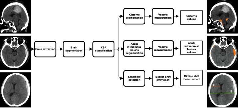
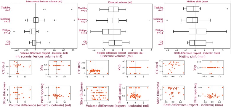
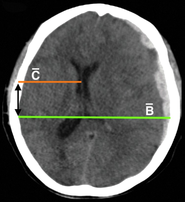
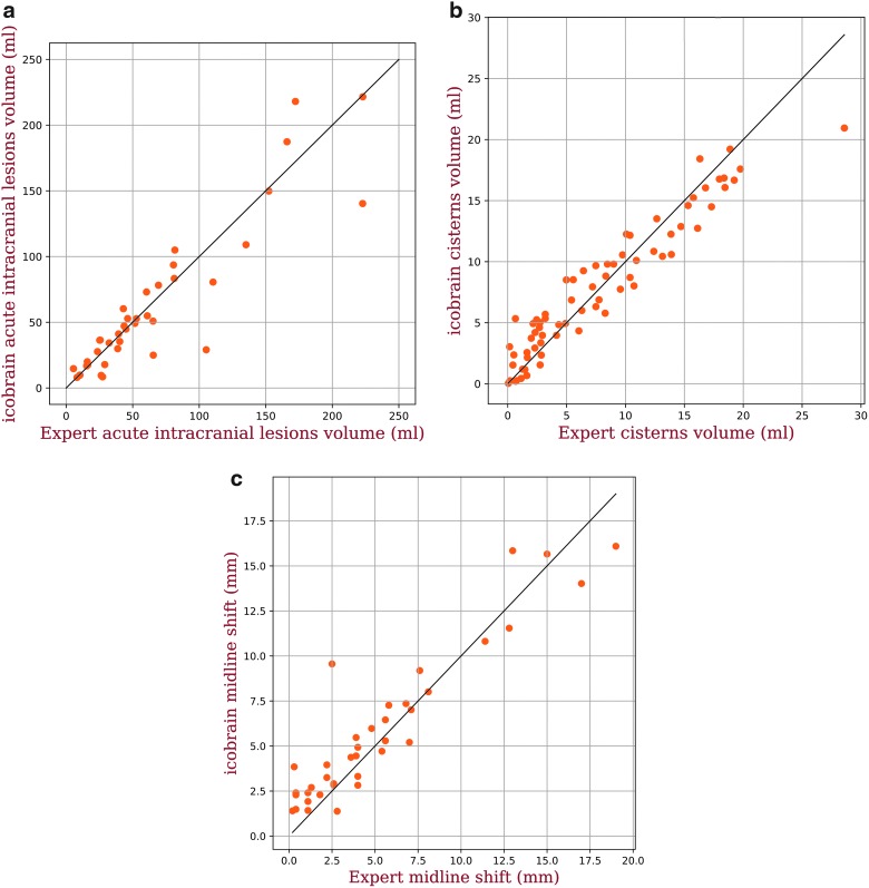
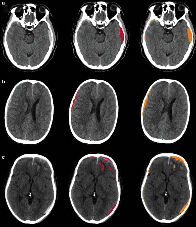
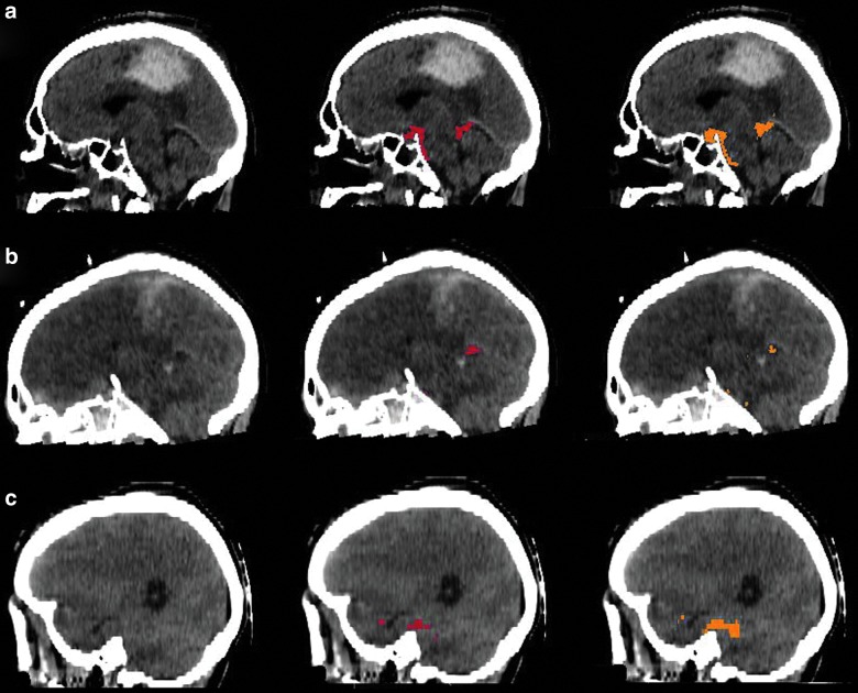
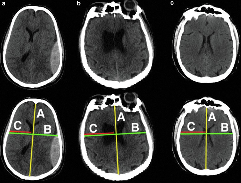
Traumatic brain injury is a complex and diverse medical condition with a high frequency of intracranial abnormalities. These can typically be visualized on a computed tomography (CT) scan, which provides important information for further patient management, such as the need for operative intervention. In order to quantify the extent of acute intracranial lesions and associated secondary injuries, such as midline shift and cisternal compression, visual assessment of CT images has limitations, including observer variability and lack of quantitative interpretation. Automated image analysis can quantify the extent of intracranial abnormalities and provide added value in routine clinical practice. In this article, we present icobrain, a fully automated method that reliably computes acute intracranial lesions volume based on deep learning, cistern volume, and midline shift on the noncontrast CT image of a patient. The accuracy of our method is evaluated on a subset of the multi-center data set from the CENTER-TBI (Collaborative European Neurotrauma Effectiveness Research in Traumatic Brain Injury) study for which expert annotations were used as a reference. Median volume differences between expert assessments and icobrain are 0.07 mL for acute intracranial lesions and -0.01 mL for cistern segmentation. Correlation between expert assessments and icobrain is 0.91 for volume of acute intracranial lesions and 0.94 for volume of the cisterns. For midline shift computations, median error is -0.22 mm, with a correlation of 0.93 with expert assessments.
创伤性脑损伤是一种复杂多样的医学病症,颅内异常的发生率很高。这些异常通常可以在计算机断层扫描(CT)上显示,为进一步的患者管理提供重要信息,例如是否需要手术干预。为了量化急性颅内病变的程度和相关的继发性损伤,如中线移位和脑池受压,CT 图像的视觉评估存在局限性,包括观察者的变异性和缺乏定量解释。自动图像分析可以量化颅内异常的程度,并在常规临床实践中提供附加价值。在本文中,我们介绍了 icobrain,这是一种完全基于深度学习的可靠计算患者非对比 CT 图像上急性颅内病变体积、脑池体积和中线移位的全自动方法。我们的方法在 CENTER-TBI(欧洲协作创伤性脑损伤有效性研究)研究的多中心数据集的一个子集上进行了评估,该数据集使用专家注释作为参考。急性颅内病变和脑池分割方面,专家评估和 icobrain 的中位数体积差异分别为 0.07 毫升和-0.01 毫升。急性颅内病变和脑池体积的专家评估和 icobrain 的相关性分别为 0.91 和 0.94。对于中线移位计算,中位误差为-0.22 毫米,与专家评估的相关性为 0.93。