• 文献检索
  • 文档翻译
  • 深度研究
  • 学术资讯
  • Suppr Zotero 插件Zotero 插件
  • 邀请有礼
  • 套餐&价格
  • 历史记录
应用&插件
Suppr Zotero 插件Zotero 插件浏览器插件Mac 客户端Windows 客户端微信小程序
定价
高级版会员购买积分包购买API积分包
服务
文献检索文档翻译深度研究API 文档MCP 服务
关于我们
关于 Suppr公司介绍联系我们用户协议隐私条款
关注我们

Suppr 超能文献

核心技术专利:CN118964589B侵权必究
粤ICP备2023148730 号-1Suppr @ 2026

文献检索

告别复杂PubMed语法,用中文像聊天一样搜索,搜遍4000万医学文献。AI智能推荐,让科研检索更轻松。

立即免费搜索

文件翻译

保留排版,准确专业,支持PDF/Word/PPT等文件格式,支持 12+语言互译。

免费翻译文档

深度研究

AI帮你快速写综述,25分钟生成高质量综述,智能提取关键信息,辅助科研写作。

立即免费体验

用于 X 射线荧光断层成像生物成像的潜在纳米粒子造影剂库。

A Library of Potential Nanoparticle Contrast Agents for X-Ray Fluorescence Tomography Bioimaging.

机构信息

Department of Applied Physics, Biomedical and X-ray Physics, KTH Royal Institute of Technology/Albanova, SE 106 91 Stockholm, Sweden.

出版信息

Contrast Media Mol Imaging. 2018 Dec 27;2018:8174820. doi: 10.1155/2018/8174820. eCollection 2018.

DOI:10.1155/2018/8174820
PMID:30686945
原文链接:https://pmc.ncbi.nlm.nih.gov/articles/PMC6327276/
Abstract

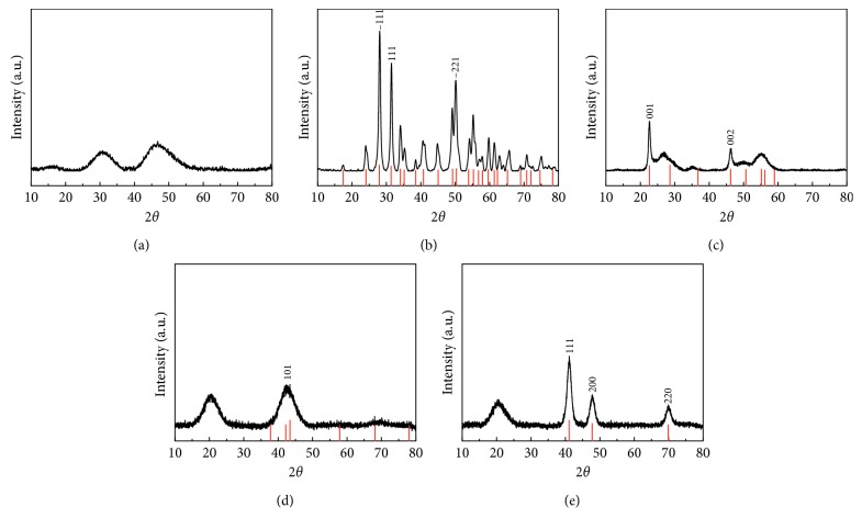
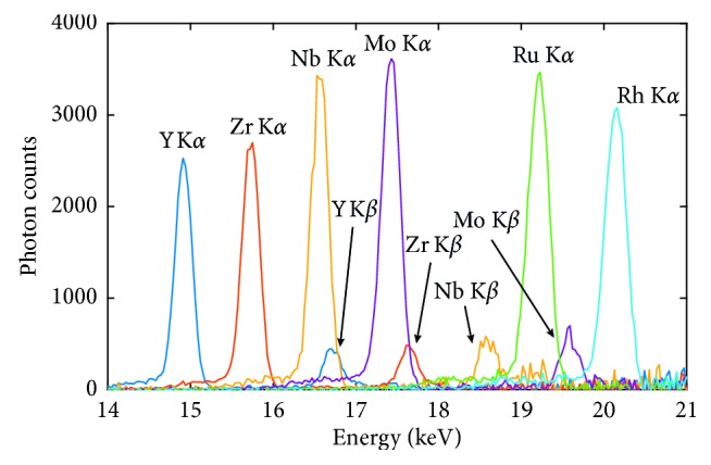
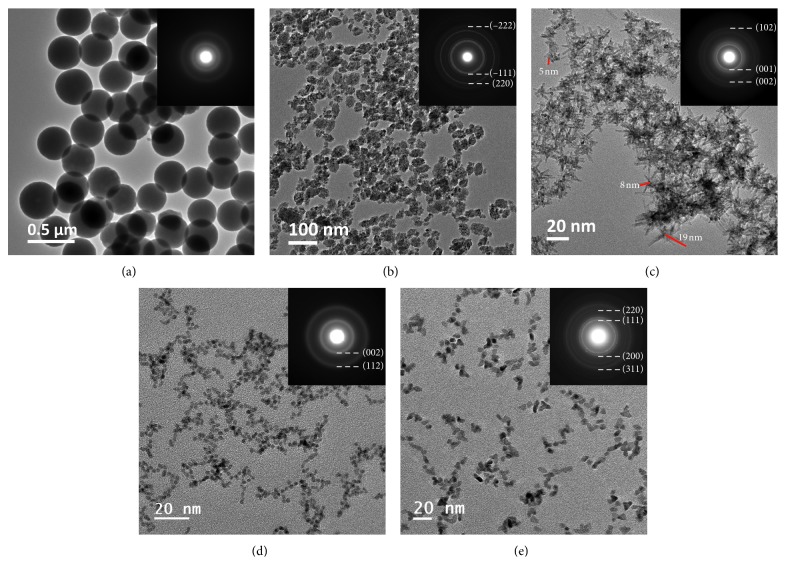
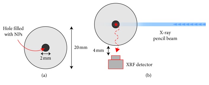
Nanoparticles (NPs) have been used as contrast agents for several bioimaging modalities. X-ray fluorescence (XRF) tomography can provide sensitive and quantitative 3D detection of NPs. With spectrally matched NPs as contrast agents, we demonstrated earlier in a laboratory system that XRF tomography could achieve high-spatial-resolution tumor imaging in mice. Here, we present the synthesis, characterization, and evaluation of a library of NPs containing Y, Zr, Nb, Rh, and Ru that have spectrally matched K-shell absorption for the laboratory scale X-ray source. The K-shell emissions of these NPs are spectrally well separated from the X-ray probe and the Compton background, making them suitable for the lab-scale XRF tomography system. Their potential as XRF contrast agents is demonstrated successfully in a small-animal equivalent phantom, confirming the simulation results. The diversity in the NP composition provides a flexible platform for a better design and biological optimization of XRF tomography nanoprobes.

摘要

纳米粒子(NPs)已被用作几种生物成像模式的对比剂。X 射线荧光(XRF)层析成像可以提供对 NPs 的敏感和定量的 3D 检测。使用光谱匹配的 NPs 作为对比剂,我们之前在实验室系统中证明,XRF 层析成像可以在小鼠中实现高空间分辨率的肿瘤成像。在这里,我们介绍了一系列含有 Y、Zr、Nb、Rh 和 Ru 的 NPs 的合成、表征和评估,这些 NPs 的 K 壳层吸收光谱与实验室规模的 X 射线源匹配。这些 NPs 的 K 壳层发射与 X 射线探针和康普顿背景光谱分离良好,使其适合实验室规模的 XRF 层析成像系统。它们在小动物等效体模中的作为 XRF 对比剂的潜力得到了成功验证,证实了模拟结果。NP 组成的多样性为 XRF 层析成像纳米探针的更好设计和生物优化提供了一个灵活的平台。

https://cdn.ncbi.nlm.nih.gov/pmc/blobs/c4ce/6327276/7f18f07698ac/CMMI2018-8174820.004.jpg
https://cdn.ncbi.nlm.nih.gov/pmc/blobs/c4ce/6327276/fdaf0331f3f8/CMMI2018-8174820.001.jpg
https://cdn.ncbi.nlm.nih.gov/pmc/blobs/c4ce/6327276/74ab59601ab4/CMMI2018-8174820.002.jpg
https://cdn.ncbi.nlm.nih.gov/pmc/blobs/c4ce/6327276/b94f21f89f80/CMMI2018-8174820.003.jpg
https://cdn.ncbi.nlm.nih.gov/pmc/blobs/c4ce/6327276/7f18f07698ac/CMMI2018-8174820.004.jpg

相似文献

1
A Library of Potential Nanoparticle Contrast Agents for X-Ray Fluorescence Tomography Bioimaging.用于 X 射线荧光断层成像生物成像的潜在纳米粒子造影剂库。
Contrast Media Mol Imaging. 2018 Dec 27;2018:8174820. doi: 10.1155/2018/8174820. eCollection 2018.
2
The feasibility of NaGdF nanoparticles as an x-ray fluorescence computed tomography imaging probe for the liver and lungs.纳米钆酸镧颗粒作为肝脏和肺部 X 射线荧光计算机断层成像探针的可行性。
Med Phys. 2020 Feb;47(2):662-671. doi: 10.1002/mp.13930. Epub 2019 Dec 10.
3
High-spatial-resolution x-ray fluorescence tomography with spectrally matched nanoparticles.高空间分辨率 X 射线荧光层析成像与光谱匹配纳米粒子。
Phys Med Biol. 2018 Aug 14;63(16):164001. doi: 10.1088/1361-6560/aad51e.
4
Quantitative imaging of gold nanoparticle distribution in a tumor-bearing mouse using benchtop x-ray fluorescence computed tomography.使用台式X射线荧光计算机断层扫描对荷瘤小鼠体内金纳米颗粒分布进行定量成像。
Sci Rep. 2016 Feb 25;6:22079. doi: 10.1038/srep22079.
5
Assessment of FLUKA, PENELOPE and MCNP6 Monte Carlo codes for estimating gold fluorescence applied to the detection of gold-infused tumoral volumes.评估用于估计金荧光以检测注入金的肿瘤体积的FLUKA、PENELOPE和MCNP6蒙特卡罗代码。
Appl Radiat Isot. 2019 Sep;151:280-288. doi: 10.1016/j.apradiso.2019.06.017. Epub 2019 Jun 12.
6
Experimental demonstration of direct L-shell x-ray fluorescence imaging of gold nanoparticles using a benchtop x-ray source.利用台式 X 射线源对金纳米粒子进行直接 L 壳层 X 射线荧光成像的实验演示。
Med Phys. 2013 Aug;40(8):080702. doi: 10.1118/1.4816297.
7
Simultaneous synthesis and amine-functionalization of single-phase BaYF5:Yb/Er nanoprobe for dual-modal in vivo upconversion fluorescence and long-lasting X-ray computed tomography imaging.单相 BaYF5:Yb/Er 纳米探针的同步合成及胺功能化用于双模态体内上转换荧光和长时 X 射线计算机断层成像。
Nanoscale. 2013 Jul 7;5(13):6023-9. doi: 10.1039/c3nr00999h. Epub 2013 May 28.
8
A Monte Carlo Model of a Benchtop X-Ray Fluorescence Computed Tomography System and Its Application to Validate a Deconvolution-Based X-Ray Fluorescence Signal Extraction Method.一种台式 X 射线荧光层析成像系统的蒙特卡罗模型及其在基于反卷积的 X 射线荧光信号提取方法验证中的应用。
IEEE Trans Med Imaging. 2018 Nov;37(11):2483-2492. doi: 10.1109/TMI.2018.2836973. Epub 2018 May 15.
9
Pinhole X-ray fluorescence imaging of gadolinium and gold nanoparticles using polychromatic X-rays: a Monte Carlo study.使用多色X射线对钆和金纳米颗粒进行针孔X射线荧光成像:一项蒙特卡罗研究。
Int J Nanomedicine. 2017 Aug 11;12:5805-5817. doi: 10.2147/IJN.S141185. eCollection 2017.
10
Quantitative Imaging of Gd Nanoparticles in Mice Using Benchtop Cone-Beam X-ray Fluorescence Computed Tomography System.使用台式锥形束 X 射线荧光计算机断层扫描系统对小鼠体内的 Gd 纳米颗粒进行定量成像。
Int J Mol Sci. 2019 May 10;20(9):2315. doi: 10.3390/ijms20092315.

引用本文的文献

1
Laboratory Liquid-Jet X-ray Microscopy and X-ray Fluorescence Imaging for Biomedical Applications.实验室液体喷射 X 射线显微镜和 X 射线荧光成像在生物医学中的应用。
Int J Mol Sci. 2024 Jan 11;25(2):920. doi: 10.3390/ijms25020920.
2
Two-Photon Polymerization Printing with High Metal Nanoparticle Loading.高金属纳米颗粒负载量的双光子聚合印刷
ACS Appl Mater Interfaces. 2023 Oct 25;15(42):49794-49804. doi: 10.1021/acsami.3c10581. Epub 2023 Oct 10.
3
Carbon Quantum Dots Conjugated Rhodium Nanoparticles as Hybrid Multimodal Contrast Agents.

本文引用的文献

1
High-spatial-resolution x-ray fluorescence tomography with spectrally matched nanoparticles.高空间分辨率 X 射线荧光层析成像与光谱匹配纳米粒子。
Phys Med Biol. 2018 Aug 14;63(16):164001. doi: 10.1088/1361-6560/aad51e.
2
Pinhole X-ray fluorescence imaging of gadolinium and gold nanoparticles using polychromatic X-rays: a Monte Carlo study.使用多色X射线对钆和金纳米颗粒进行针孔X射线荧光成像:一项蒙特卡罗研究。
Int J Nanomedicine. 2017 Aug 11;12:5805-5817. doi: 10.2147/IJN.S141185. eCollection 2017.
3
Quantitative imaging of gold nanoparticle distribution in a tumor-bearing mouse using benchtop x-ray fluorescence computed tomography.
碳量子点共轭铑纳米颗粒作为混合多模态造影剂
Nanomaterials (Basel). 2021 Aug 24;11(9):2165. doi: 10.3390/nano11092165.
4
Optical and X-ray Fluorescent Nanoparticles for Dual Mode Bioimaging.用于双模生物成像的光学和 X 射线荧光纳米颗粒。
ACS Nano. 2021 Mar 23;15(3):5077-5085. doi: 10.1021/acsnano.0c10127. Epub 2021 Feb 15.
5
Synthesis, Physicochemical Characterization, and Cytotoxicity Assessment of Rh Nanoparticles with Different Morphologies-as Potential XFCT Nanoprobes.不同形态铑纳米颗粒作为潜在X射线荧光计算机断层扫描纳米探针的合成、物理化学表征及细胞毒性评估
Nanomaterials (Basel). 2020 Oct 27;10(11):2129. doi: 10.3390/nano10112129.
6
Synthesis and Cytotoxicity Studies on Ru and Rh Nanoparticles as Potential X-Ray Fluorescence Computed Tomography (XFCT) Contrast Agents.钌和铑纳米颗粒作为潜在的X射线荧光计算机断层扫描(XFCT)造影剂的合成及细胞毒性研究
Nanomaterials (Basel). 2020 Feb 12;10(2):310. doi: 10.3390/nano10020310.
使用台式X射线荧光计算机断层扫描对荷瘤小鼠体内金纳米颗粒分布进行定量成像。
Sci Rep. 2016 Feb 25;6:22079. doi: 10.1038/srep22079.
4
Laboratory x-ray fluorescence tomography for high-resolution nanoparticle bio-imaging.用于高分辨率纳米颗粒生物成像的实验室X射线荧光断层扫描技术
Opt Lett. 2014 May 1;39(9):2790-3. doi: 10.1364/OL.39.002790.
5
Use of portable X-ray fluorescence (PXRF) in vivo as an alternative technique for the assessment of iron levels in patients with thalassemia and hemochromatosis.便携式 X 射线荧光(PXRF)在体内作为评估地中海贫血和血色病患者铁水平的替代技术的应用。
Health Phys. 2013 Feb;104(2):132-8. doi: 10.1097/HP.0b013e3182667721.
6
Experimental demonstration of benchtop x-ray fluorescence computed tomography (XFCT) of gold nanoparticle-loaded objects using lead- and tin-filtered polychromatic cone-beams.使用铅和锡过滤的多色锥形束对负载金纳米粒子的物体进行台式 X 射线荧光计算机层析成像(XFCT)的实验演示。
Phys Med Biol. 2012 Dec 7;57(23):N457-67. doi: 10.1088/0031-9155/57/23/N457. Epub 2012 Nov 8.
7
An early investigation of ytterbium nanocolloids for selective and quantitative "multicolor" spectral CT imaging.镱纳米胶体用于选择性和定量“多色”光谱 CT 成像的早期研究。
ACS Nano. 2012 Apr 24;6(4):3364-70. doi: 10.1021/nn300392x. Epub 2012 Mar 8.
8
Nanoparticles as computed tomography contrast agents: current status and future perspectives.纳米粒子作为计算机断层扫描造影剂:现状与展望。
Nanomedicine (Lond). 2012 Feb;7(2):257-69. doi: 10.2217/nnm.11.190.
9
Magnetic nanoparticles for cancer diagnosis and therapy.用于癌症诊断和治疗的磁性纳米颗粒。
Pharm Res. 2012 May;29(5):1180-8. doi: 10.1007/s11095-012-0679-7. Epub 2012 Jan 25.
10
Multifunctional nanoprobes for upconversion fluorescence, MR and CT trimodal imaging.多功能上转换荧光/MR/CT 三模态成像探针。
Biomaterials. 2012 Feb;33(4):1079-89. doi: 10.1016/j.biomaterials.2011.10.039. Epub 2011 Nov 5.