Research Center for Advanced Science and Technology, The University of Tokyo, Japan.
Department of Neurosurgery, Graduate School of Medicine, The University of Tokyo, Japan.
Neuroimage Clin. 2019;22:101684. doi: 10.1016/j.nicl.2019.101684. Epub 2019 Jan 22.
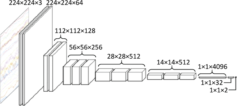
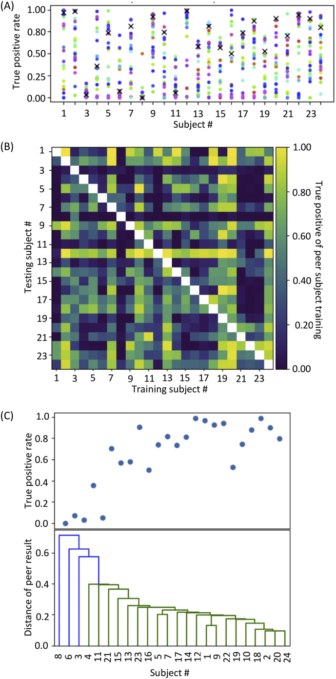
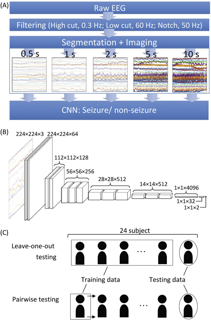
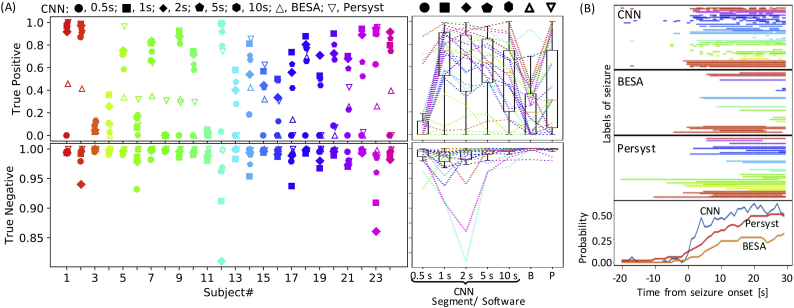
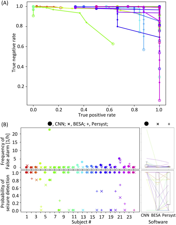
We hypothesized that expert epileptologists can detect seizures directly by visually analyzing EEG plot images, unlike automated methods that analyze spectro-temporal features or complex, non-stationary features of EEG signals. If so, seizure detection could benefit from convolutional neural networks because their visual recognition ability is comparable to that of humans. We explored image-based seizure detection by applying convolutional neural networks to long-term EEG that included epileptic seizures. After filtering, EEG data were divided into short segments based on a given time window and converted into plot EEG images, each of which was classified by convolutional neural networks as 'seizure' or 'non-seizure'. These resultant labels were then used to design a clinically practical index for seizure detection. The best true positive rate was obtained using a 1-s time window. The median true positive rate of convolutional neural networks labelling by seconds was 74%, which was higher than that of commercially available seizure detection software (20% by BESA and 31% by Persyst). For practical use, the median of detected seizure rate by minutes was 100% by convolutional neural networks, which was higher than the 73.3% by BESA and 81.7% by Persyst. The false alarm of convolutional neural networks' seizure detection was issued at 0.2 per hour, which appears acceptable for clinical practice. Moreover, we demonstrated that seizure detection improved when training was performed using EEG patterns similar to those of testing data, suggesting that adding a variety of seizure patterns to the training dataset will improve our method. Thus, artificial visual recognition by convolutional neural networks allows for seizure detection, which otherwise currently relies on skillful visual inspection by expert epileptologists during clinical diagnosis.
我们假设,与分析 EEG 信号的时频特征或复杂、非平稳特征的自动化方法不同,专家癫痫学家可以通过直观地分析 EEG 图谱图像来直接检测癫痫发作。如果是这样,那么癫痫发作的检测可能会受益于卷积神经网络,因为它们的视觉识别能力与人类相当。我们通过将卷积神经网络应用于包含癫痫发作的长期 EEG 来探索基于图像的癫痫发作检测。经过滤波后,根据给定的时间窗口将 EEG 数据分为短段,并将其转换为图谱 EEG 图像,每个图像都由卷积神经网络分类为“癫痫发作”或“非癫痫发作”。然后,这些得到的标签被用于设计一种用于癫痫发作检测的临床实用指标。使用 1 秒的时间窗口可以获得最佳的真阳性率。卷积神经网络以秒为单位标记的中位数真阳性率为 74%,高于商用癫痫发作检测软件(BESA 为 20%,Persyst 为 31%)。为了实际应用,卷积神经网络以分钟为单位检测到的癫痫发作率的中位数为 100%,高于 BESA 的 73.3%和 Persyst 的 81.7%。卷积神经网络的癫痫发作检测假阳性率为每小时 0.2 次,这似乎可以接受。此外,我们还证明,使用与测试数据相似的 EEG 模式进行训练可以提高癫痫发作检测的效果,这表明向训练数据集添加各种癫痫发作模式将改进我们的方法。因此,卷积神经网络的人工视觉识别可以实现癫痫发作检测,而目前这种检测依赖于专家癫痫学家在临床诊断中进行熟练的视觉检查。