Department of Physiology & Membrane Biology, University of California Davis, Davis, CA, USA.
Department of Pharmacology, University of California Davis, Davis, CA, USA.
J Physiol. 2019 Apr;597(8):2139-2162. doi: 10.1113/JP277283. Epub 2019 Mar 12.
Prevailing dogma holds that activation of the β-adrenergic receptor/cAMP/protein kinase A signalling pathway leads to enhanced L-type Ca 1.2 channel activity, resulting in increased Ca influx into ventricular myocytes and a positive inotropic response. However, the full mechanistic and molecular details underlying this phenomenon are incompletely understood. Ca 1.2 channel clusters decorate T-tubule sarcolemmas of ventricular myocytes. Within clusters, nanometer proximity between channels permits Ca -dependent co-operative gating behaviour mediated by physical interactions between adjacent channel C-terminal tails. We report that stimulation of cardiomyocytes with isoproterenol, evokes dynamic, protein kinase A-dependent augmentation of Ca 1.2 channel abundance along cardiomyocyte T-tubules, resulting in the appearance of channel 'super-clusters', and enhanced channel co-operativity that amplifies Ca influx. On the basis of these data, we suggest a new model in which a sub-sarcolemmal pool of pre-synthesized Ca 1.2 channels resides in cardiomyocytes and can be mobilized to the membrane in times of high haemodynamic or metabolic demand, to tune excitation-contraction coupling.
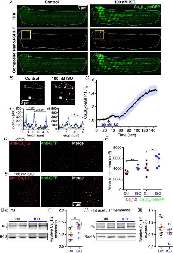
Voltage-dependent L-type Ca 1.2 channels play an indispensable role in cardiac excitation-contraction coupling. Activation of the β-adrenergic receptor (βAR)/cAMP/protein kinase A (PKA) signalling pathway leads to enhanced Ca 1.2 activity, resulting in increased Ca influx into ventricular myocytes and a positive inotropic response. Ca 1.2 channels exhibit a clustered distribution along the T-tubule sarcolemma of ventricular myocytes where nanometer proximity between channels permits Ca -dependent co-operative gating behaviour mediated by dynamic, physical, allosteric interactions between adjacent channel C-terminal tails. This amplifies Ca influx and augments myocyte Ca transient and contraction amplitudes. We investigated whether βAR signalling could alter Ca 1.2 channel clustering to facilitate co-operative channel interactions and elevate Ca influx in ventricular myocytes. Bimolecular fluorescence complementation experiments reveal that the βAR agonist, isoproterenol (ISO), promotes enhanced Ca 1.2-Ca 1.2 physical interactions. Super-resolution nanoscopy and dynamic channel tracking indicate that these interactions are expedited by enhanced spatial proximity between channels, resulting in the appearance of Ca 1.2 'super-clusters' along the z-lines of ISO-stimulated cardiomyocytes. The mechanism that leads to super-cluster formation involves rapid, dynamic augmentation of sarcolemmal Ca 1.2 channel abundance after ISO application. Optical and electrophysiological single channel recordings confirm that these newly inserted channels are functional and contribute to overt co-operative gating behaviour of Ca 1.2 channels in ISO stimulated myocytes. The results of the present study reveal a new facet of βAR-mediated regulation of Ca 1.2 channels in the heart and support the novel concept that a pre-synthesized pool of sub-sarcolemmal Ca 1.2 channel-containing vesicles/endosomes resides in cardiomyocytes and can be mobilized to the sarcolemma to tune excitation-contraction coupling to meet metabolic and/or haemodynamic demands.
流行的观点认为,β-肾上腺素能受体/cAMP/蛋白激酶 A 信号通路的激活导致 L 型 Ca 1.2 通道活性增强,导致心室肌细胞内 Ca 流入增加和正性变力反应。然而,这种现象的完整机制和分子细节尚不完全清楚。Ca 1.2 通道簇装饰心室肌细胞的 T 小管肌膜。在簇内,通道之间的纳米级接近允许通过相邻通道 C 末端尾部之间的物理相互作用介导的 Ca 依赖性协同门控行为。我们报告说,用异丙肾上腺素刺激心肌细胞会引起 Ca 1.2 通道丰度沿心肌细胞 T 小管的动态、蛋白激酶 A 依赖性增强,导致通道“超簇”的出现,并增强通道协同性,从而增加 Ca 流入。基于这些数据,我们提出了一个新的模型,即在心肌细胞中存在预先合成的 Ca 1.2 通道的亚肌膜池,并且可以在高血流动力学或代谢需求时动员到膜上,以调节兴奋-收缩偶联。
电压依赖性 L 型 Ca 1.2 通道在心脏兴奋-收缩偶联中起着不可或缺的作用。β-肾上腺素能受体(βAR)/cAMP/蛋白激酶 A(PKA)信号通路的激活导致 Ca 1.2 活性增强,导致心室肌细胞内 Ca 流入增加和正性变力反应。Ca 1.2 通道沿心室肌细胞的 T 小管肌膜呈簇状分布,通道之间的纳米级接近允许通过相邻通道 C 末端尾部之间的动态、物理、变构相互作用介导 Ca 依赖性协同门控行为。这放大了 Ca 流入并增强了心肌细胞 Ca 瞬变和收缩幅度。我们研究了 βAR 信号是否可以改变 Ca 1.2 通道簇集以促进协同通道相互作用并增加心室肌细胞中的 Ca 流入。双分子荧光互补实验表明,βAR 激动剂异丙肾上腺素(ISO)促进增强的 Ca 1.2-Ca 1.2 物理相互作用。超分辨率纳米显微镜和动态通道跟踪表明,这些相互作用通过通道之间空间接近度的增加而加速,导致 ISO 刺激的心肌细胞沿 z 线出现 Ca 1.2“超簇”。导致超簇形成的机制涉及 ISO 应用后肌膜 Ca 1.2 通道丰度的快速动态增强。光学和电生理单通道记录证实,这些新插入的通道是功能性的,并有助于 ISO 刺激的肌细胞中 Ca 1.2 通道的明显协同门控行为。本研究的结果揭示了 βAR 介导的心脏 Ca 1.2 通道调节的一个新方面,并支持一个新的概念,即在心肌细胞中存在预先合成的亚肌膜 Ca 1.2 通道含囊泡/内体池,可以动员到肌膜以调节兴奋-收缩偶联以满足代谢和/或血流动力学需求。