• 文献检索
  • 文档翻译
  • 深度研究
  • 学术资讯
  • Suppr Zotero 插件Zotero 插件
  • 邀请有礼
  • 套餐&价格
  • 历史记录
应用&插件
Suppr Zotero 插件Zotero 插件浏览器插件Mac 客户端Windows 客户端微信小程序
定价
高级版会员购买积分包购买API积分包
服务
文献检索文档翻译深度研究API 文档MCP 服务
关于我们
关于 Suppr公司介绍联系我们用户协议隐私条款
关注我们

Suppr 超能文献

核心技术专利:CN118964589B侵权必究
粤ICP备2023148730 号-1Suppr @ 2026

文献检索

告别复杂PubMed语法,用中文像聊天一样搜索,搜遍4000万医学文献。AI智能推荐,让科研检索更轻松。

立即免费搜索

文件翻译

保留排版,准确专业,支持PDF/Word/PPT等文件格式,支持 12+语言互译。

免费翻译文档

深度研究

AI帮你快速写综述,25分钟生成高质量综述,智能提取关键信息,辅助科研写作。

立即免费体验

基于人羊膜的组织工程皮肤替代物的研制。

Development of a tissue-engineered skin substitute on a base of human amniotic membrane.

作者信息

John Samuel, Kesting Marco Rainer, Paulitschke Philipp, Stöckelhuber Mechthild, von Bomhard Achim

机构信息

University Hospital Jena, Jena, Germany.

Department of Oral and Maxillofacial Surgery, Technical University of Munich, Munich, Germany.

出版信息

J Tissue Eng. 2019 Feb 2;10:2041731418825378. doi: 10.1177/2041731418825378. eCollection 2019 Jan-Dec.

DOI:10.1177/2041731418825378
PMID:30746119
原文链接:https://pmc.ncbi.nlm.nih.gov/articles/PMC6360641/
Abstract

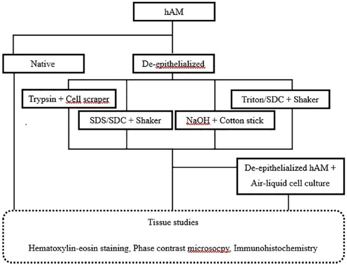
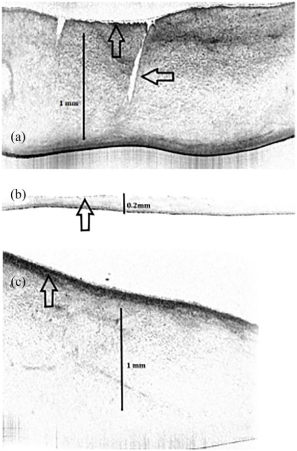
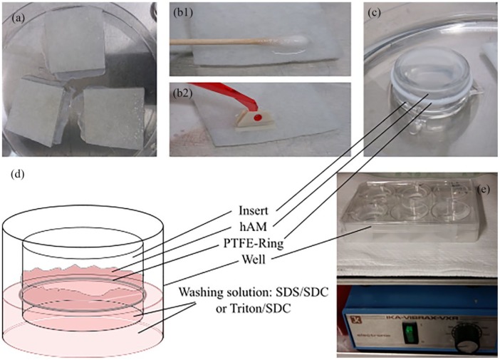
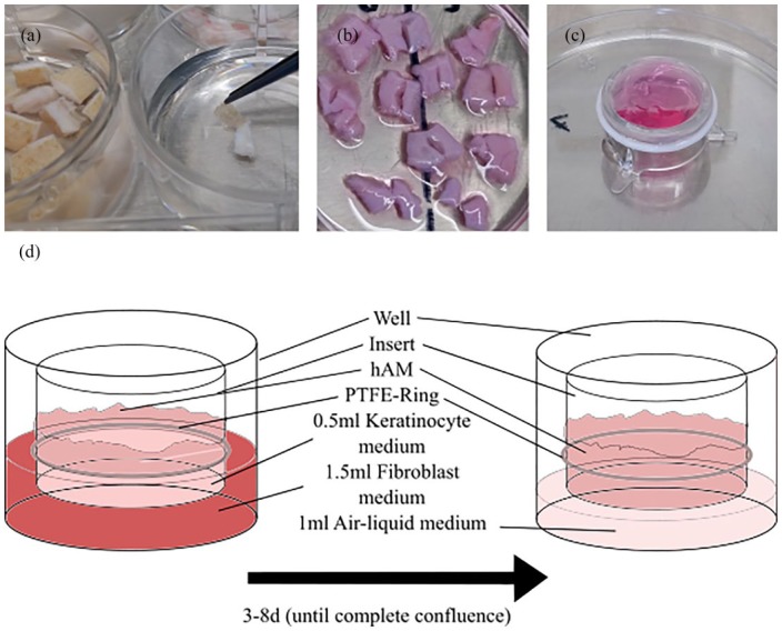
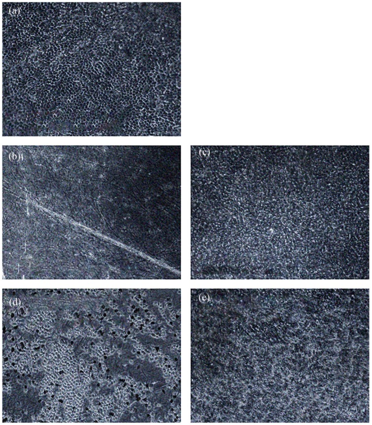
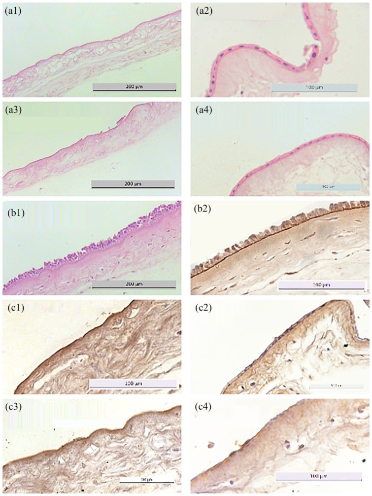
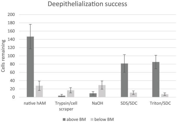
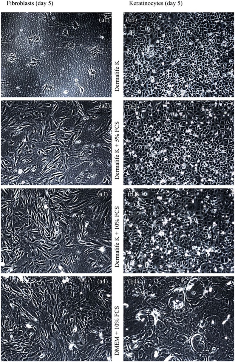
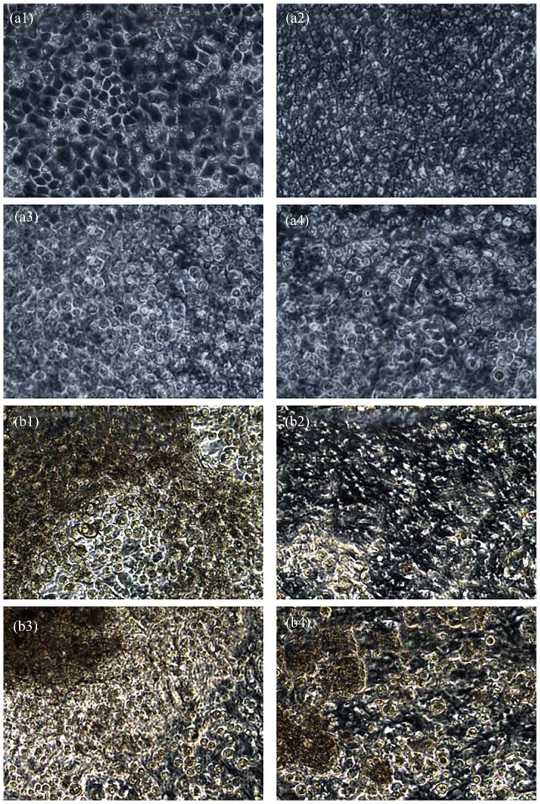
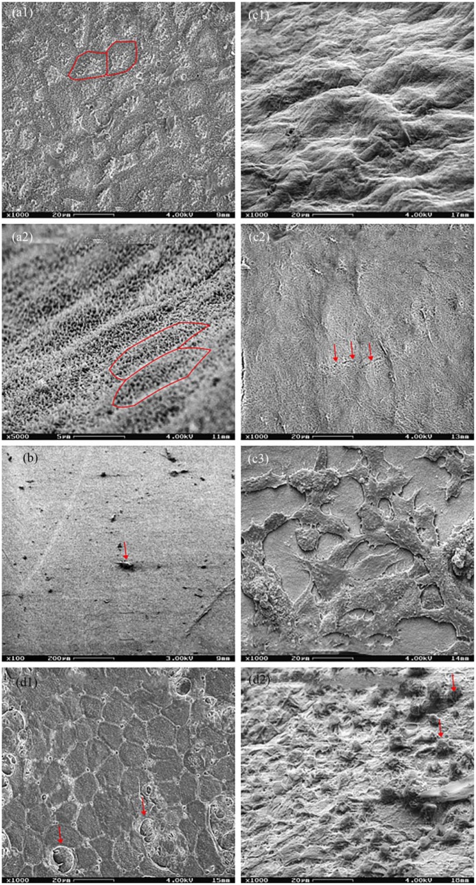
Allogenic graft material and tissue engineering have recently shown promising results for the improvement of both esthetic and functional outcomes in the treatment of large skin defects. We chose human amniotic membrane as a cellular scaffold in order to develop a skin substitute for later in vivo uses. Various methods of de-epithelialization of the human amniotic membrane were evaluated by histological analysis including hematoxylin-eosin and laminin staining, optic coherence tomography, and scanning electron microscopy with 0.25/0.02% trypsin/ethylenediaminetetraacetic acid treatment and mechanical cell removal showing an almost complete loss of the epithelium and a mainly intact basement membrane. Novel examination of human amniotic membrane by optic coherence tomography was feasible, but difficulties were experienced in handling and interpretation of the tissue as no comparable data exist. Subsequently, we developed an air-liquid interface cell culture to cultivate keratinocytes and fibroblasts on the de-epithelialized human amniotic membrane. We achieved a mostly keratinized surface on the epidermal side with a confluent fibroblast network on the chorion side.

摘要

同种异体移植材料和组织工程最近在治疗大面积皮肤缺损方面,在改善美学和功能效果上都显示出了令人鼓舞的结果。我们选择人羊膜作为细胞支架,以便开发一种皮肤替代物供日后体内使用。通过组织学分析评估了人羊膜去上皮化的各种方法,包括苏木精-伊红和层粘连蛋白染色、光学相干断层扫描以及用0.25/0.02%胰蛋白酶/乙二胺四乙酸处理和机械去除细胞的扫描电子显微镜检查,结果显示上皮几乎完全丧失,基底膜基本完整。用光学相干断层扫描对人羊膜进行新的检查是可行的,但由于没有可比数据,在处理和解释组织时遇到了困难。随后,我们开发了气液界面细胞培养法,在去上皮化的人羊膜上培养角质形成细胞和成纤维细胞。我们在表皮侧实现了大部分角质化表面,在绒毛膜侧形成了融合的成纤维细胞网络。

https://cdn.ncbi.nlm.nih.gov/pmc/blobs/5f69/6360641/3a57c689ff99/10.1177_2041731418825378-fig10.jpg
https://cdn.ncbi.nlm.nih.gov/pmc/blobs/5f69/6360641/7b7aa3c9dcd9/10.1177_2041731418825378-fig1.jpg
https://cdn.ncbi.nlm.nih.gov/pmc/blobs/5f69/6360641/121bc6db7bcd/10.1177_2041731418825378-fig2.jpg
https://cdn.ncbi.nlm.nih.gov/pmc/blobs/5f69/6360641/bb75ccf2cbed/10.1177_2041731418825378-fig3.jpg
https://cdn.ncbi.nlm.nih.gov/pmc/blobs/5f69/6360641/061947e93fc8/10.1177_2041731418825378-fig4.jpg
https://cdn.ncbi.nlm.nih.gov/pmc/blobs/5f69/6360641/7b49acc24ce6/10.1177_2041731418825378-fig5.jpg
https://cdn.ncbi.nlm.nih.gov/pmc/blobs/5f69/6360641/b7db8a81c5bd/10.1177_2041731418825378-fig6.jpg
https://cdn.ncbi.nlm.nih.gov/pmc/blobs/5f69/6360641/21ba02679ecf/10.1177_2041731418825378-fig7.jpg
https://cdn.ncbi.nlm.nih.gov/pmc/blobs/5f69/6360641/c0b0dca663c2/10.1177_2041731418825378-fig8.jpg
https://cdn.ncbi.nlm.nih.gov/pmc/blobs/5f69/6360641/1863c7e3be33/10.1177_2041731418825378-fig9.jpg
https://cdn.ncbi.nlm.nih.gov/pmc/blobs/5f69/6360641/3a57c689ff99/10.1177_2041731418825378-fig10.jpg

相似文献

1
Development of a tissue-engineered skin substitute on a base of human amniotic membrane.基于人羊膜的组织工程皮肤替代物的研制。
J Tissue Eng. 2019 Feb 2;10:2041731418825378. doi: 10.1177/2041731418825378. eCollection 2019 Jan-Dec.
2
The enzymatic de-epithelialization technique determines denuded amniotic membrane integrity and viability of harvested epithelial cells.酶去上皮化技术决定了采集的上皮细胞的无上皮羊膜完整性和活力。
PLoS One. 2018 Mar 27;13(3):e0194820. doi: 10.1371/journal.pone.0194820. eCollection 2018.
3
A skin substitute based on human amniotic membrane.一种基于人羊膜的皮肤替代物。
Cell Tissue Bank. 2014 Jun;15(2):257-65. doi: 10.1007/s10561-014-9427-z. Epub 2014 Feb 16.
4
Using human epithelial amnion cells in human de-epidermized dermis for skin regeneration.将人羊膜上皮细胞用于人去表皮真皮以实现皮肤再生。
J Dermatol Sci. 2016 Jan;81(1):26-34. doi: 10.1016/j.jdermsci.2015.10.018. Epub 2015 Oct 31.
5
Acellular amniotic membrane: an appropriate scaffold for fibroblast proliferation.脱细胞羊膜:成纤维细胞增殖的合适支架。
Clin Exp Dermatol. 2013 Aug;38(6):646-51. doi: 10.1111/ced.12087.
6
Evaluation of Tissue-engineered Skin on Base of Human Amniotic Membrane for Wound Healing.基于人羊膜的组织工程皮肤用于伤口愈合的评估
Plast Reconstr Surg Glob Open. 2019 Jul 25;7(7):e2320. doi: 10.1097/GOX.0000000000002320. eCollection 2019 Jul.
7
Urea-De-Epithelialized Human Amniotic Membrane for Ocular Surface Reconstruction.去上皮化人尿囊膜在眼表重建中的应用。
Stem Cells Transl Med. 2019 Jul;8(7):620-626. doi: 10.1002/sctm.18-0201. Epub 2019 Mar 13.
8
Fabrication and characterization of spongy denuded amniotic membrane based scaffold for tissue engineering.海绵去表皮羊膜基支架的制备及性能表征用于组织工程。
Cell J. 2015 Winter;16(4):476-87. doi: 10.22074/cellj.2015.493. Epub 2015 Jan 13.
9
Supportive properties of basement membrane layer of human amniotic membrane enable development of tissue engineering applications.人羊膜基底膜层的支持特性有助于组织工程应用的发展。
Cell Tissue Bank. 2018 Sep;19(3):357-371. doi: 10.1007/s10561-017-9680-z. Epub 2018 Jan 8.
10
Cultured corneal epithelia for ocular surface disease.用于眼表疾病的培养角膜上皮细胞
Trans Am Ophthalmol Soc. 1999;97:891-986.

引用本文的文献

1
Sterilization of Human Amniotic Membrane Using an Ozone Hydrodynamic System.臭氧流体动力学系统对人羊膜的消毒。
Ann Biomed Eng. 2024 May;52(5):1425-1434. doi: 10.1007/s10439-024-03467-3. Epub 2024 Feb 27.
2
Application of Amniotic Membrane in Skin Regeneration.羊膜在皮肤再生中的应用。
Pharmaceutics. 2023 Feb 23;15(3):748. doi: 10.3390/pharmaceutics15030748.
3
Comparison of human amniotic membrane decellularisation approaches for hESC-derived RPE cells culture.比较人羊膜脱细胞方法用于 hESC 衍生的 RPE 细胞培养。

本文引用的文献

1
Gamma-irradiated human amniotic membrane decellularised with sodium dodecyl sulfate is a more efficient substrate for the ex vivo expansion of limbal stem cells.经伽马射线辐照并用十二烷基硫酸钠脱细胞化的人羊膜是体外扩增角膜缘干细胞的更有效底物。
Acta Biomater. 2017 Oct 1;61:124-133. doi: 10.1016/j.actbio.2017.07.041. Epub 2017 Jul 29.
2
An Ultra-thin Amniotic Membrane as Carrier in Corneal Epithelium Tissue-Engineering.超薄羊膜作为角膜上皮组织工程的载体
Sci Rep. 2016 Feb 15;6:21021. doi: 10.1038/srep21021.
3
Trypan Blue Exclusion Test of Cell Viability.
BMJ Open Ophthalmol. 2022 Sep;7(1). doi: 10.1136/bmjophth-2022-000981.
4
Recent update on craniofacial tissue engineering.颅面组织工程学的最新进展
J Tissue Eng. 2021 Apr 20;12:20417314211003735. doi: 10.1177/20417314211003735. eCollection 2021 Jan-Dec.
5
Bioengineering Vascular Networks to Study Angiogenesis and Vascularization of Physiologically Relevant Tissue Models .生物工程血管网络以研究生理相关组织模型中的血管生成和血管化。
ACS Biomater Sci Eng. 2020 Jun 8;6(6):3513-3528. doi: 10.1021/acsbiomaterials.0c00191. Epub 2020 Apr 29.
6
Evaluation of Radiosterilized Glyercerolated Amniotic Membranes as a Substrate for Cultured Human Epithelial Cells.评估辐照甘油化羊膜作为培养人上皮细胞的基质。
Organogenesis. 2020;16(1):27-41. doi: 10.1080/15476278.2020.1723366. Epub 2020 Feb 15.
7
Regulating Preparation Of Functional Alginate-Chitosan Three-Dimensional Scaffold For Skin Tissue Engineering.调控用于皮肤组织工程的功能性藻酸盐-壳聚糖三维支架的制备。
Int J Nanomedicine. 2019 Nov 13;14:8891-8903. doi: 10.2147/IJN.S210329. eCollection 2019.
8
Evaluation of Tissue-engineered Skin on Base of Human Amniotic Membrane for Wound Healing.基于人羊膜的组织工程皮肤用于伤口愈合的评估
Plast Reconstr Surg Glob Open. 2019 Jul 25;7(7):e2320. doi: 10.1097/GOX.0000000000002320. eCollection 2019 Jul.
细胞活力的台盼蓝排斥试验
Curr Protoc Immunol. 2015 Nov 2;111:A3.B.1-A3.B.3. doi: 10.1002/0471142735.ima03bs111.
4
Amniotic membrane: new concepts for an old dressing.羊膜:一种古老敷料的新概念。
Wound Repair Regen. 2014 Jul-Aug;22(4):451-6. doi: 10.1111/wrr.12188.
5
Evaluation of human amniotic membrane as a wound dressing for split-thickness skin-graft donor sites.人羊膜作为中厚皮片供皮区伤口敷料的评估。
Biomed Res Int. 2014;2014:572183. doi: 10.1155/2014/572183. Epub 2014 Jun 9.
6
Manual hematoxylin and eosin staining of mouse tissue sections.小鼠组织切片的苏木精和伊红手工染色。
Cold Spring Harb Protoc. 2014 Jun 2;2014(6):655-8. doi: 10.1101/pdb.prot073411.
7
Use of synthetic serum-free medium for culture of human dermal fibroblasts to establish an experimental system similar to living dermis.使用合成无血清培养基培养人真皮成纤维细胞,建立类似于活体真皮的实验系统。
Cytotechnology. 2015 May;67(3):507-14. doi: 10.1007/s10616-014-9709-0. Epub 2014 Mar 1.
8
Immunohistochemical expression of basement membrane laminin in histological grades of oral squamous cell carcinoma: A semiquantitative analysis.口腔鳞状细胞癌组织学分级中基底膜层粘连蛋白的免疫组织化学表达:一项半定量分析。
J Oral Maxillofac Pathol. 2013 May;17(2):185-9. doi: 10.4103/0973-029X.119755.
9
A simple alkaline method for decellularizing human amniotic membrane for cell culture.一种用于细胞培养的人羊膜脱细胞的简单碱性方法。
PLoS One. 2013 Nov 13;8(11):e79632. doi: 10.1371/journal.pone.0079632. eCollection 2013.
10
Culturing of human nasal epithelial cells at the air liquid interface.人鼻上皮细胞在气液界面的培养。
J Vis Exp. 2013 Oct 8(80):50646. doi: 10.3791/50646.