Department of Physics, King's College London, London, United Kingdom.
Randall Centre for Cell and Molecular Biophysics, King's College London, London, United Kingdom.
PLoS One. 2019 Feb 14;14(2):e0211165. doi: 10.1371/journal.pone.0211165. eCollection 2019.
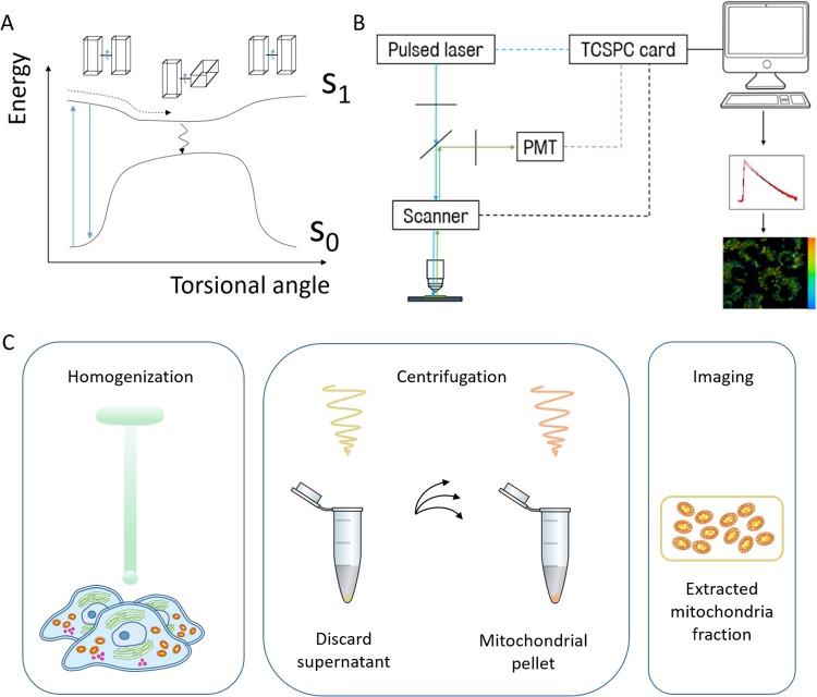
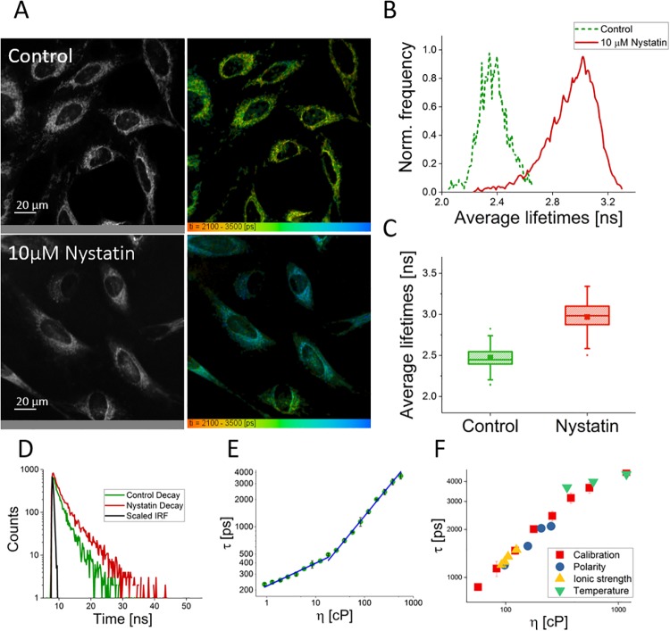
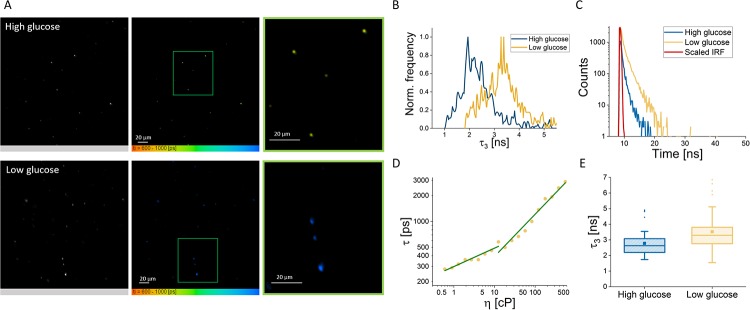
The only way to visually observe cellular viscosity, which can greatly influence biological reactions and has been linked to several human diseases, is through viscosity imaging. Imaging cellular viscosity has allowed the mapping of viscosity in cells, and the next frontier is targeted viscosity imaging of organelles and their microenvironments. Here we present a fluorescent molecular rotor/FLIM framework to image both organellar viscosity and membrane fluidity, using a combination of chemical targeting and organelle extraction. For demonstration, we image matrix viscosity and membrane fluidity of mitochondria, which have been linked to human diseases, including Alzheimer's Disease and Leigh's syndrome. We find that both are highly dynamic and responsive to small environmental and physiological changes, even under non-pathological conditions. This shows that neither viscosity nor fluidity can be assumed to be fixed and underlines the need for single-cell, and now even single-organelle, imaging.
观察细胞粘度的唯一方法是通过粘度成像,细胞粘度会极大地影响生物反应,并且与几种人类疾病有关。粘度成像可以绘制细胞内的粘度图,下一个前沿领域是靶向细胞器及其微环境的粘度成像。在这里,我们提出了一种荧光分子转子/FLIM 框架,使用化学靶向和细胞器提取的组合来成像细胞器的粘度和膜流动性。为了演示,我们对与人类疾病(包括阿尔茨海默病和 Leigh 综合征)有关的线粒体的基质粘度和膜流动性进行成像。我们发现,即使在非病理条件下,两者都是高度动态的,并对小的环境和生理变化做出反应。这表明,无论是粘度还是流动性都不能被认为是固定的,这强调了单细胞,甚至是单细胞成像的必要性。