• 文献检索
  • 文档翻译
  • 深度研究
  • 学术资讯
  • Suppr Zotero 插件Zotero 插件
  • 邀请有礼
  • 套餐&价格
  • 历史记录
应用&插件
Suppr Zotero 插件Zotero 插件浏览器插件Mac 客户端Windows 客户端微信小程序
定价
高级版会员购买积分包购买API积分包
服务
文献检索文档翻译深度研究API 文档MCP 服务
关于我们
关于 Suppr公司介绍联系我们用户协议隐私条款
关注我们

Suppr 超能文献

核心技术专利:CN118964589B侵权必究
粤ICP备2023148730 号-1Suppr @ 2026

文献检索

告别复杂PubMed语法,用中文像聊天一样搜索,搜遍4000万医学文献。AI智能推荐,让科研检索更轻松。

立即免费搜索

文件翻译

保留排版,准确专业,支持PDF/Word/PPT等文件格式,支持 12+语言互译。

免费翻译文档

深度研究

AI帮你快速写综述,25分钟生成高质量综述,智能提取关键信息,辅助科研写作。

立即免费体验

乳腺磁共振筛查项目中纤维-腺体组织(FGT)和背景实质强化(BPE)与乳腺癌风险和假阳性的关系:一项回顾性队列研究。

Amount of fibroglandular tissue FGT and background parenchymal enhancement BPE in relation to breast cancer risk and false positives in a breast MRI screening program : A retrospective cohort study.

机构信息

Department of Radiology and Nuclear Medicine, Radboud University Medical Center, Geert Grooteplein 10, route 766, 6525 GA, Nijmegen, the Netherlands.

Department of Pathology, Radboud University Medical Center, Nijmegen, the Netherlands.

出版信息

Eur Radiol. 2019 Sep;29(9):4678-4690. doi: 10.1007/s00330-019-06020-2. Epub 2019 Feb 22.

DOI:10.1007/s00330-019-06020-2
PMID:30796568
原文链接:https://pmc.ncbi.nlm.nih.gov/articles/PMC6682856/
Abstract

OBJECTIVES

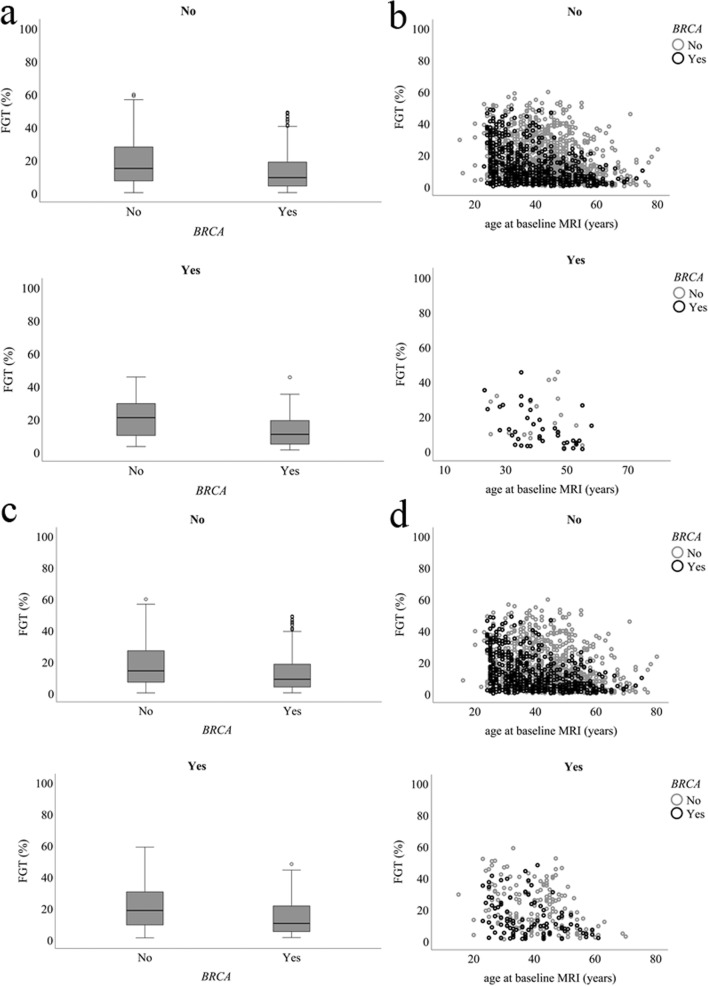
The purpose of this study is to evaluate the predictive value of the amount of fibroglandular tissue (FGT) and background parenchymal enhancement (BPE), measured at baseline on breast MRI, for breast cancer development and risk of false-positive findings in women at increased risk for breast cancer.

METHODS

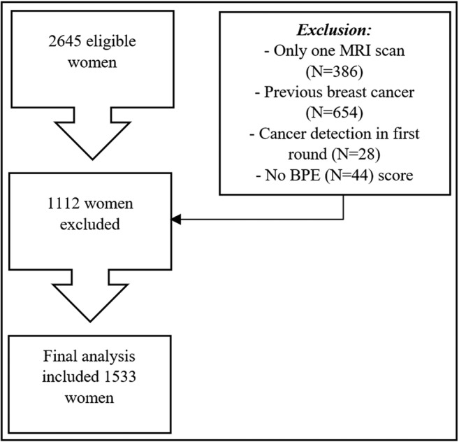
Negative baseline MRI scans of 1533 women participating in a screening program for women at increased risk for breast cancer between January 1, 2003, and January 1, 2014, were selected. Automated tools based on deep learning were used to obtain quantitative measures of FGT and BPE. Logistic regression using forward selection was used to assess relationships between FGT, BPE, cancer detection, false-positive recall, and false-positive biopsy.

RESULTS

Sixty cancers were detected in follow-up. FGT was only associated to short-term cancer risk; BPE was not associated with cancer risk. High FGT and BPE did lead to more false-positive recalls at baseline (OR 1.259, p = 0.050, and OR 1.475, p = 0.003) and to more frequent false-positive biopsies at baseline (OR 1.315, p = 0.049, and OR 1.807, p = 0.002), but were not predictive for false-positive findings in subsequent screening rounds.

CONCLUSIONS

FGT and BPE, measured on baseline MRI, are not predictive for overall breast cancer development in women at increased risk. High FGT and BPE lead to more false-positive findings at baseline.

KEY POINTS

• Amount of fibroglandular tissue is only predictive for short-term breast cancer risk in women at increased risk. • Background parenchymal enhancement measured on baseline MRI is not predictive for breast cancer development in women at increased risk. • High amount of fibroglandular tissue and background parenchymal enhancement lead to more false-positive findings at baseline MRI.

摘要

目的

本研究旨在评估基线期乳腺 MRI 上测量的纤维腺体组织(FGT)和背景实质强化(BPE)的量对乳腺癌发展和乳腺癌高风险女性假阳性发现的风险的预测价值。

方法

选取 2003 年 1 月 1 日至 2014 年 1 月 1 日期间参加乳腺癌高风险筛查计划的 1533 名女性的阴性基线 MRI 扫描。使用基于深度学习的自动工具获得 FGT 和 BPE 的定量测量值。使用向前选择的逻辑回归评估 FGT、BPE、癌症检出、假阳性召回和假阳性活检之间的关系。

结果

在随访中检测到 60 例癌症。FGT 仅与短期癌症风险相关;BPE 与癌症风险无关。高 FGT 和 BPE 确实导致基线期更多的假阳性召回(OR 1.259,p=0.050 和 OR 1.475,p=0.003)和更多的基线期假阳性活检(OR 1.315,p=0.049 和 OR 1.807,p=0.002),但不能预测后续筛查轮次的假阳性发现。

结论

基线期 MRI 上测量的 FGT 和 BPE 不能预测乳腺癌高风险女性的总体乳腺癌发展。高 FGT 和 BPE 导致基线期更多的假阳性发现。

关键点

• 纤维腺体组织的量仅可预测乳腺癌高风险女性的短期乳腺癌风险。• 基线期 MRI 上测量的背景实质强化不能预测乳腺癌高风险女性的乳腺癌发展。• 高纤维腺体组织量和背景实质强化导致基线期 MRI 上更多的假阳性发现。

https://cdn.ncbi.nlm.nih.gov/pmc/blobs/d8d6/6682856/13aa7573743c/330_2019_6020_Fig4_HTML.jpg
https://cdn.ncbi.nlm.nih.gov/pmc/blobs/d8d6/6682856/9148594305bd/330_2019_6020_Fig1_HTML.jpg
https://cdn.ncbi.nlm.nih.gov/pmc/blobs/d8d6/6682856/f4dac4639c90/330_2019_6020_Fig2_HTML.jpg
https://cdn.ncbi.nlm.nih.gov/pmc/blobs/d8d6/6682856/0a31d6135e06/330_2019_6020_Fig3_HTML.jpg
https://cdn.ncbi.nlm.nih.gov/pmc/blobs/d8d6/6682856/13aa7573743c/330_2019_6020_Fig4_HTML.jpg

相似文献

1
Amount of fibroglandular tissue FGT and background parenchymal enhancement BPE in relation to breast cancer risk and false positives in a breast MRI screening program : A retrospective cohort study.乳腺磁共振筛查项目中纤维-腺体组织(FGT)和背景实质强化(BPE)与乳腺癌风险和假阳性的关系:一项回顾性队列研究。
Eur Radiol. 2019 Sep;29(9):4678-4690. doi: 10.1007/s00330-019-06020-2. Epub 2019 Feb 22.
2
Background parenchymal enhancement and fibroglandular tissue on breast MRI in women with high genetic risk: Are changes before and after risk-reducing salpingo-oophorectomy associated with breast cancer risk?背景实质强化和乳腺磁共振成像中高遗传风险女性的纤维腺体组织:降低风险的输卵管卵巢切除术前后的变化是否与乳腺癌风险相关?
Eur J Radiol. 2018 Dec;109:171-177. doi: 10.1016/j.ejrad.2018.10.030. Epub 2018 Nov 2.
3
The impact of bilateral salpingo-oophorectomy on breast MRI background parenchymal enhancement and fibroglandular tissue.双侧输卵管卵巢切除术对乳腺 MRI 背景实质增强和纤维腺体组织的影响。
Eur Radiol. 2014 Jan;24(1):162-8. doi: 10.1007/s00330-013-2993-9. Epub 2013 Aug 28.
4
MRI Background Parenchymal Enhancement Is Not Associated with Breast Cancer.MRI背景实质强化与乳腺癌无关。
PLoS One. 2016 Jul 5;11(7):e0158573. doi: 10.1371/journal.pone.0158573. eCollection 2016.
5
Quantitative Assessment of Breast Parenchymal Uptake on 18F-FDG PET/CT: Correlation with Age, Background Parenchymal Enhancement, and Amount of Fibroglandular Tissue on MRI.18F-FDG PET/CT对乳腺实质摄取的定量评估:与年龄、背景实质强化及MRI上纤维腺体量的相关性
J Nucl Med. 2016 Oct;57(10):1518-1522. doi: 10.2967/jnumed.116.174904. Epub 2016 May 26.
6
Quantitative assessment of background parenchymal enhancement in breast MRI predicts response to risk-reducing salpingo-oophorectomy: preliminary evaluation in a cohort of BRCA1/2 mutation carriers.乳腺MRI中背景实质强化的定量评估可预测降低风险的输卵管卵巢切除术的反应:BRCA1/2突变携带者队列的初步评估
Breast Cancer Res. 2015 May 19;17:67. doi: 10.1186/s13058-015-0577-0.
7
Impact of menopausal status on background parenchymal enhancement and fibroglandular tissue on breast MRI.绝经状态对乳腺 MRI 背景实质强化和纤维腺体组织的影响。
Eur Radiol. 2012 Dec;22(12):2641-7. doi: 10.1007/s00330-012-2553-8. Epub 2012 Jul 4.
8
Histopathologic characteristics of background parenchymal enhancement (BPE) on breast MRI.乳腺 MRI 背景实质强化(BPE)的组织病理学特征。
Breast Cancer Res Treat. 2018 Nov;172(2):487-496. doi: 10.1007/s10549-018-4916-6. Epub 2018 Aug 23.
9
Impact of tamoxifen on amount of fibroglandular tissue, background parenchymal enhancement, and cysts on breast magnetic resonance imaging.他莫昔芬对乳腺磁共振成像中纤维腺体组织、背景实质强化和囊肿数量的影响。
Breast J. 2012 Nov-Dec;18(6):527-34. doi: 10.1111/tbj.12002. Epub 2012 Sep 25.
10
Fully automatic quantification of fibroglandular tissue and background parenchymal enhancement with accurate implementation for axial and sagittal breast MRI protocols.轴向和矢状位乳腺 MRI 方案中准确实施的纤维腺体组织和背景实质增强的全自动定量。
Med Phys. 2021 Jan;48(1):238-252. doi: 10.1002/mp.14581. Epub 2020 Nov 27.

引用本文的文献

1
Using artificial intelligence system for assisting the classification of breast ultrasound glandular tissue components in dense breast tissue.使用人工智能系统辅助对致密乳腺组织中的乳腺超声腺体组织成分进行分类。
Sci Rep. 2025 Apr 6;15(1):11754. doi: 10.1038/s41598-025-95871-5.
2
Impact of menopause and age on breast density and background parenchymal enhancement in dynamic contrast-enhanced magnetic resonance imaging.绝经和年龄对动态对比增强磁共振成像中乳腺密度和背景实质强化的影响。
J Med Imaging (Bellingham). 2025 Nov;12(Suppl 2):S22002. doi: 10.1117/1.JMI.12.S2.S22002. Epub 2025 Mar 11.
3
Breast MRI to Screen Women With Extremely Dense Breasts.

本文引用的文献

1
Influence of Risk Category and Screening Round on the Performance of an MR Imaging and Mammography Screening Program in Carriers of the BRCA Mutation and Other Women at Increased Risk.BRCA 基因突变携带者和其他高风险女性的风险类别和筛查轮次对磁共振成像和乳房 X 线筛查项目性能的影响。
Radiology. 2018 Feb;286(2):443-451. doi: 10.1148/radiol.2017170458. Epub 2017 Oct 16.
2
Complementary Value of Contralateral Parenchymal Enhancement on DCE-MRI to Prognostic Models and Molecular Assays in High-risk ER/HER2 Breast Cancer.DCE-MRI 对侧实质增强对高危 ER/HER2 乳腺癌预后模型和分子检测的补充价值。
Clin Cancer Res. 2017 Nov 1;23(21):6505-6515. doi: 10.1158/1078-0432.CCR-17-0176. Epub 2017 Aug 8.
3
乳腺磁共振成像用于筛查乳腺极度致密的女性。
J Magn Reson Imaging. 2025 Jul;62(1):58-72. doi: 10.1002/jmri.29716. Epub 2025 Jan 24.
4
Machine Learning Assessment of Background Parenchymal Enhancement in Breast Cancer and Clinical Applications: A Literature Review.乳腺癌背景实质强化的机器学习评估及临床应用:文献综述
Cancers (Basel). 2024 Oct 31;16(21):3681. doi: 10.3390/cancers16213681.
5
How Does Diagnostic Accuracy Evolve with Increased Breast MRI Experience?随着乳腺 MRI 经验的增加,诊断准确性如何演变?
Tomography. 2023 Nov 6;9(6):2067-2078. doi: 10.3390/tomography9060162.
6
Quantitative evaluation of Kaiser score in diagnosing breast dynamic contrast-enhanced magnetic resonance imaging for patients with high-grade background parenchymal enhancement.凯泽评分在诊断背景实质强化程度较高患者乳腺动态对比增强磁共振成像中的定量评估
Quant Imaging Med Surg. 2023 Oct 1;13(10):6384-6394. doi: 10.21037/qims-23-113. Epub 2023 Aug 17.
7
Association of Breast Cancer Odds with Background Parenchymal Enhancement Quantified Using a Fully Automated Method at MRI: The IMAGINE Study.基于 MRI 的全自动背景实质增强量化评估与乳腺癌患病风险的相关性:IMAGINE 研究。
Radiology. 2023 Sep;308(3):e230367. doi: 10.1148/radiol.230367.
8
The Role of Diffusion-Weighted Imaging Based on Maximum-Intensity Projection in Young Patients with Marked Background Parenchymal Enhancement on Contrast-Enhanced Breast MRI.基于最大强度投影的扩散加权成像在乳腺对比增强磁共振成像中背景实质明显强化的年轻患者中的作用
Life (Basel). 2023 Aug 14;13(8):1744. doi: 10.3390/life13081744.
9
Beyond Breast Density: Risk Measures for Breast Cancer in Multiple Imaging Modalities.超越乳腺密度:多种影像学模式下乳腺癌的风险评估指标。
Radiology. 2023 Mar;306(3):e222575. doi: 10.1148/radiol.222575. Epub 2023 Feb 7.
10
The Frequency and Causes of Not-Detected Breast Malignancy in Dynamic Contrast-Enhanced MRI.动态对比增强磁共振成像中未检测到的乳腺恶性肿瘤的频率及原因
Diagnostics (Basel). 2022 Oct 24;12(11):2575. doi: 10.3390/diagnostics12112575.
Breast cancer risk models: a comprehensive overview of existing models, validation, and clinical applications.
乳腺癌风险模型:现有模型的全面概述、验证及临床应用。
Breast Cancer Res Treat. 2017 Jul;164(2):263-284. doi: 10.1007/s10549-017-4247-z. Epub 2017 Apr 25.
4
Using deep learning to segment breast and fibroglandular tissue in MRI volumes.利用深度学习对磁共振成像(MRI)容积中的乳腺和纤维腺组织进行分割。
Med Phys. 2017 Feb;44(2):533-546. doi: 10.1002/mp.12079.
5
Volumetric breast density affects performance of digital screening mammography.乳腺体积密度会影响数字化乳腺筛查钼靶摄影的效果。
Breast Cancer Res Treat. 2017 Feb;162(1):95-103. doi: 10.1007/s10549-016-4090-7. Epub 2016 Dec 23.
6
Reply to "Breast MRI background parenchymal enhancement (BPE) correlates with the risk of breast cancer".对《乳腺磁共振成像背景实质强化(BPE)与乳腺癌风险相关》的回复
Magn Reson Imaging. 2016 Nov;34(9):1337-1338. doi: 10.1016/j.mri.2016.07.013. Epub 2016 Jul 27.
7
MRI Background Parenchymal Enhancement Is Not Associated with Breast Cancer.MRI背景实质强化与乳腺癌无关。
PLoS One. 2016 Jul 5;11(7):e0158573. doi: 10.1371/journal.pone.0158573. eCollection 2016.
8
Breast MRI background parenchymal enhancement (BPE) correlates with the risk of breast cancer.乳腺磁共振成像背景实质强化(BPE)与乳腺癌风险相关。
Magn Reson Imaging. 2016 Feb;34(2):173-6. doi: 10.1016/j.mri.2015.10.014. Epub 2015 Oct 24.
9
Quantitative assessment of background parenchymal enhancement in breast MRI predicts response to risk-reducing salpingo-oophorectomy: preliminary evaluation in a cohort of BRCA1/2 mutation carriers.乳腺MRI中背景实质强化的定量评估可预测降低风险的输卵管卵巢切除术的反应:BRCA1/2突变携带者队列的初步评估
Breast Cancer Res. 2015 May 19;17:67. doi: 10.1186/s13058-015-0577-0.
10
Are Qualitative Assessments of Background Parenchymal Enhancement, Amount of Fibroglandular Tissue on MR Images, and Mammographic Density Associated with Breast Cancer Risk?磁共振成像中背景实质强化、纤维腺体量的定性评估以及乳腺X线密度与乳腺癌风险相关吗?
Radiology. 2015 Aug;276(2):371-80. doi: 10.1148/radiol.2015142304. Epub 2015 May 12.