• 文献检索
  • 文档翻译
  • 深度研究
  • 学术资讯
  • Suppr Zotero 插件Zotero 插件
  • 邀请有礼
  • 套餐&价格
  • 历史记录
应用&插件
Suppr Zotero 插件Zotero 插件浏览器插件Mac 客户端Windows 客户端微信小程序
定价
高级版会员购买积分包购买API积分包
服务
文献检索文档翻译深度研究API 文档MCP 服务
关于我们
关于 Suppr公司介绍联系我们用户协议隐私条款
关注我们

Suppr 超能文献

核心技术专利:CN118964589B侵权必究
粤ICP备2023148730 号-1Suppr @ 2026

文献检索

告别复杂PubMed语法,用中文像聊天一样搜索,搜遍4000万医学文献。AI智能推荐,让科研检索更轻松。

立即免费搜索

文件翻译

保留排版,准确专业,支持PDF/Word/PPT等文件格式,支持 12+语言互译。

免费翻译文档

深度研究

AI帮你快速写综述,25分钟生成高质量综述,智能提取关键信息,辅助科研写作。

立即免费体验

从 CT 扫描图像中检测肺结节的有效且可靠框架。

Effective and Reliable Framework for Lung Nodules Detection from CT Scan Images.

机构信息

Department of Software Engineering, Foundation University Islamabad, Islamabad, Pakistan.

Department of Computer Science, Shaheed Zulfikar Ali Bhutto Institute of Science and Technology, Islamabad, Pakistan.

出版信息

Sci Rep. 2019 Mar 21;9(1):4989. doi: 10.1038/s41598-019-41510-9.

DOI:10.1038/s41598-019-41510-9
PMID:30899052
原文链接:https://pmc.ncbi.nlm.nih.gov/articles/PMC6428823/
Abstract

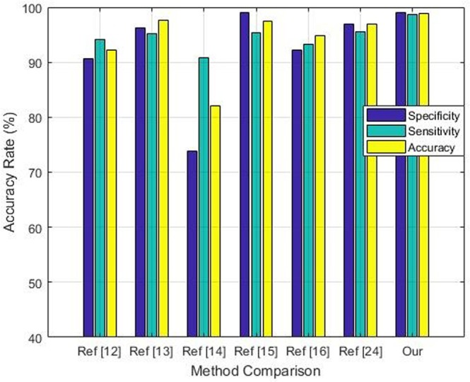
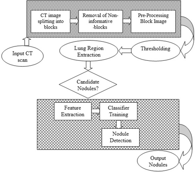
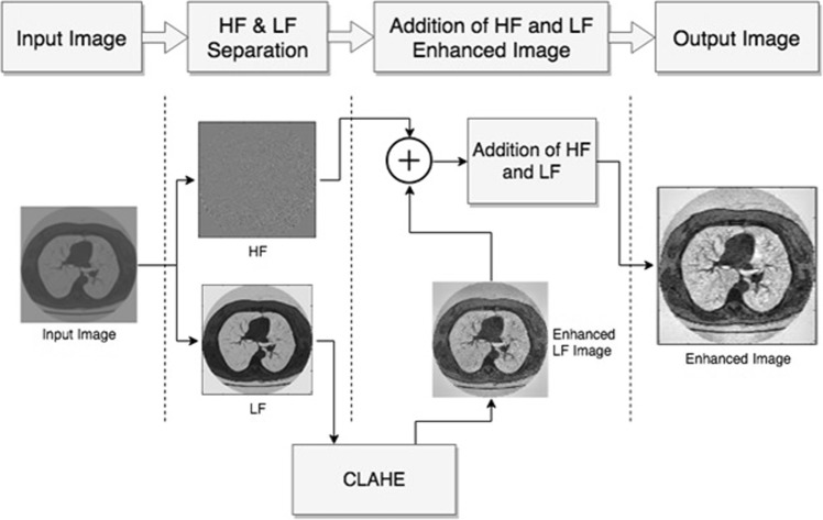
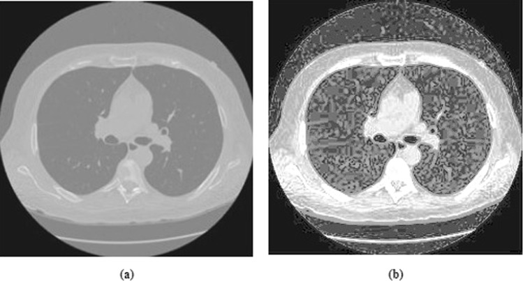
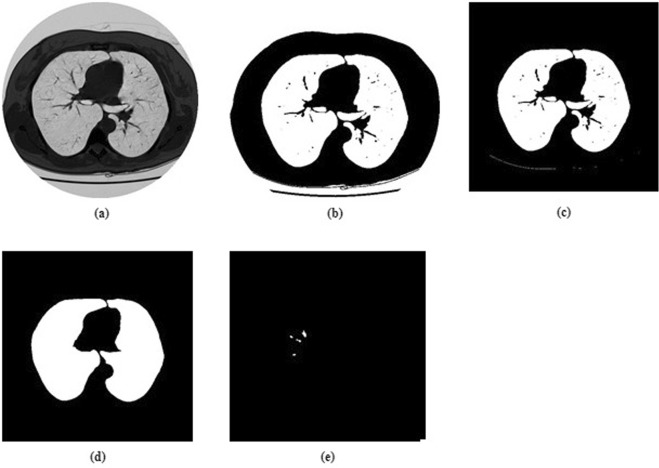
Lung cancer is considered more serious among other prevailing cancer types. One of the reasons for it is that it is usually not diagnosed until it has spread and by that time it becomes very difficult to treat. Early detection of lung cancer can significantly increase the chances of survival of a cancer patient. An effective nodule detection system can play a key role in early detection of lung cancer thus increasing the chances of successful treatment. In this research work, we have proposed a novel classification framework for nodule classification. The framework consists of multiple phases that include image contrast enhancement, segmentation, optimal feature extraction, followed by employment of these features for training and testing of Support Vector Machine. We have empirically tested the efficacy of our technique by utilizing the well-known Lung Image Consortium Database (LIDC) dataset. The empirical results suggest that the technique is highly effective for reducing the false positive rates. We were able to receive an impressive sensitivity rate of 97.45%.

摘要

肺癌在其他常见癌症类型中被认为更为严重。其中一个原因是,通常在肺癌扩散后才被诊断出来,而此时治疗难度很大。早期发现肺癌可以显著提高癌症患者的生存机会。一个有效的结节检测系统可以在肺癌的早期检测中发挥关键作用,从而提高成功治疗的机会。在这项研究工作中,我们提出了一种新的结节分类框架。该框架由多个阶段组成,包括图像对比度增强、分割、最优特征提取,然后利用这些特征进行支持向量机的训练和测试。我们通过利用著名的肺部图像联盟数据库 (LIDC) 数据集,对我们的技术的有效性进行了实证测试。实验结果表明,该技术对降低假阳性率非常有效。我们能够获得令人印象深刻的 97.45%的灵敏度。

https://cdn.ncbi.nlm.nih.gov/pmc/blobs/92f2/6428823/dbfdf9be8266/41598_2019_41510_Fig13_HTML.jpg
https://cdn.ncbi.nlm.nih.gov/pmc/blobs/92f2/6428823/77f9bcff4593/41598_2019_41510_Fig1_HTML.jpg
https://cdn.ncbi.nlm.nih.gov/pmc/blobs/92f2/6428823/f2e24f33e1d3/41598_2019_41510_Fig2_HTML.jpg
https://cdn.ncbi.nlm.nih.gov/pmc/blobs/92f2/6428823/17f509fb424c/41598_2019_41510_Fig3_HTML.jpg
https://cdn.ncbi.nlm.nih.gov/pmc/blobs/92f2/6428823/dfe0be457da5/41598_2019_41510_Fig4_HTML.jpg
https://cdn.ncbi.nlm.nih.gov/pmc/blobs/92f2/6428823/c3d7ba93c4ae/41598_2019_41510_Fig5_HTML.jpg
https://cdn.ncbi.nlm.nih.gov/pmc/blobs/92f2/6428823/6f35dd242a91/41598_2019_41510_Fig6_HTML.jpg
https://cdn.ncbi.nlm.nih.gov/pmc/blobs/92f2/6428823/40c3f8c5e9b9/41598_2019_41510_Fig7_HTML.jpg
https://cdn.ncbi.nlm.nih.gov/pmc/blobs/92f2/6428823/8c0bd1a89366/41598_2019_41510_Fig8_HTML.jpg
https://cdn.ncbi.nlm.nih.gov/pmc/blobs/92f2/6428823/fc267ac6d4db/41598_2019_41510_Fig9_HTML.jpg
https://cdn.ncbi.nlm.nih.gov/pmc/blobs/92f2/6428823/e5d59fc1ba2b/41598_2019_41510_Fig10_HTML.jpg
https://cdn.ncbi.nlm.nih.gov/pmc/blobs/92f2/6428823/2ba5cb114c93/41598_2019_41510_Fig11_HTML.jpg
https://cdn.ncbi.nlm.nih.gov/pmc/blobs/92f2/6428823/2755eb2813b3/41598_2019_41510_Fig12_HTML.jpg
https://cdn.ncbi.nlm.nih.gov/pmc/blobs/92f2/6428823/dbfdf9be8266/41598_2019_41510_Fig13_HTML.jpg

相似文献

1
Effective and Reliable Framework for Lung Nodules Detection from CT Scan Images.从 CT 扫描图像中检测肺结节的有效且可靠框架。
Sci Rep. 2019 Mar 21;9(1):4989. doi: 10.1038/s41598-019-41510-9.
2
Lungs nodule detection framework from computed tomography images using support vector machine.基于支持向量机的 CT 图像肺部结节检测框架。
Microsc Res Tech. 2019 Aug;82(8):1256-1266. doi: 10.1002/jemt.23275. Epub 2019 Apr 11.
3
Lung nodule segmentation and recognition using SVM classifier and active contour modeling: a complete intelligent system.使用 SVM 分类器和主动轮廓建模进行肺结节分割和识别:一个完整的智能系统。
Comput Biol Med. 2013 May;43(4):287-300. doi: 10.1016/j.compbiomed.2012.12.004. Epub 2013 Jan 29.
4
Fully automatic detection of lung nodules in CT images using a hybrid feature set.利用混合特征集自动检测 CT 图像中的肺结节。
Med Phys. 2017 Jul;44(7):3615-3629. doi: 10.1002/mp.12273. Epub 2017 Jun 16.
5
An adaptive morphology based segmentation technique for lung nodule detection in thoracic CT image.一种基于自适应形态学的胸部CT图像肺结节检测分割技术。
Comput Methods Programs Biomed. 2020 Dec;197:105720. doi: 10.1016/j.cmpb.2020.105720. Epub 2020 Aug 25.
6
Multistage segmentation model and SVM-ensemble for precise lung nodule detection.多阶段分割模型和 SVM 集成用于精确肺结节检测。
Int J Comput Assist Radiol Surg. 2018 Jul;13(7):1083-1095. doi: 10.1007/s11548-018-1715-9. Epub 2018 Feb 28.
7
A Segmentation Framework of Pulmonary Nodules in Lung CT Images.肺部CT图像中肺结节的分割框架
J Digit Imaging. 2016 Feb;29(1):86-103. doi: 10.1007/s10278-015-9801-9.
8
A weighted rule based method for predicting malignancy of pulmonary nodules by nodule characteristics.一种基于加权规则的方法,通过结节特征预测肺结节的恶性程度。
J Biomed Inform. 2015 Aug;56:69-79. doi: 10.1016/j.jbi.2015.05.011. Epub 2015 May 22.
9
A new computationally efficient CAD system for pulmonary nodule detection in CT imagery.一种新的计算效率高的 CT 图像肺结节检测 CAD 系统。
Med Image Anal. 2010 Jun;14(3):390-406. doi: 10.1016/j.media.2010.02.004. Epub 2010 Feb 19.
10
Automatic lung nodule detection using multi-scale dot nodule-enhancement filter and weighted support vector machines in chest computed tomography.基于多尺度点结节增强滤波器和加权支持向量机的胸部 CT 自动肺结节检测。
PLoS One. 2019 Jan 10;14(1):e0210551. doi: 10.1371/journal.pone.0210551. eCollection 2019.

引用本文的文献

1
ROI extraction in corona virus (COVID 19) CT images using intuitionistic fuzzy edge detection.基于直觉模糊边缘检测的冠状病毒(COVID-19)CT图像中的感兴趣区域提取
Heliyon. 2024 Mar 16;10(6):e27798. doi: 10.1016/j.heliyon.2024.e27798. eCollection 2024 Mar 30.
2
Diagnostic Accuracy of Machine Learning AI Architectures in Detection and Classification of Lung Cancer: A Systematic Review.机器学习人工智能架构在肺癌检测与分类中的诊断准确性:一项系统综述。
Diagnostics (Basel). 2023 Jun 22;13(13):2145. doi: 10.3390/diagnostics13132145.
3
A radiomics approach for lung nodule detection in thoracic CT images based on the dynamic patterns of morphological variation.

本文引用的文献

1
Multistage segmentation model and SVM-ensemble for precise lung nodule detection.多阶段分割模型和 SVM 集成用于精确肺结节检测。
Int J Comput Assist Radiol Surg. 2018 Jul;13(7):1083-1095. doi: 10.1007/s11548-018-1715-9. Epub 2018 Feb 28.
2
An approach for reducing the error rate in automated lung segmentation.一种降低自动肺部分割错误率的方法。
Comput Biol Med. 2016 Sep 1;76:143-53. doi: 10.1016/j.compbiomed.2016.06.022. Epub 2016 Jun 29.
3
Automated system for lung nodules classification based on wavelet feature descriptor and support vector machine.
基于形态变化动态模式的胸部 CT 图像肺结节检测的放射组学方法。
Eur Radiol. 2022 Jun;32(6):3767-3777. doi: 10.1007/s00330-021-08456-x. Epub 2022 Jan 12.
4
A Novel Pulmonary Nodule Detection Model Based on Multi-Step Cascaded Networks.基于多步级联网络的新型肺结节检测模型。
Sensors (Basel). 2020 Aug 1;20(15):4301. doi: 10.3390/s20154301.
基于小波特征描述符和支持向量机的肺结节自动分类系统
Biomed Eng Online. 2015 Feb 12;14:9. doi: 10.1186/s12938-015-0003-y.
4
Shape "break-and-repair" strategy and its application to automated medical image segmentation.形状“断裂-修复”策略及其在医学图像自动分割中的应用。
IEEE Trans Vis Comput Graph. 2011 Jan;17(1):115-24. doi: 10.1109/TVCG.2010.56.
5
WLD: a robust local image descriptor.WLD:一种强大的局部图像描述符。
IEEE Trans Pattern Anal Mach Intell. 2010 Sep;32(9):1705-20. doi: 10.1109/TPAMI.2009.155.
6
Random forest based lung nodule classification aided by clustering.基于聚类的随机森林肺结节分类辅助方法。
Comput Med Imaging Graph. 2010 Oct;34(7):535-42. doi: 10.1016/j.compmedimag.2010.03.006. Epub 2010 Apr 28.
7
Toward automated segmentation of the pathological lung in CT.迈向CT图像中病理性肺部的自动分割
IEEE Trans Med Imaging. 2005 Aug;24(8):1025-38. doi: 10.1109/TMI.2005.851757.
8
Informatics in radiology (infoRAD): new tools for computer assistance in thoracic CT. Part 1. Functional analysis of lungs, lung lobes, and bronchopulmonary segments.放射学中的信息学(infoRAD):胸部CT计算机辅助的新工具。第1部分。肺、肺叶和支气管肺段的功能分析。
Radiographics. 2005 Mar-Apr;25(2):525-36. doi: 10.1148/rg.252045070.
9
Lung image database consortium: developing a resource for the medical imaging research community.肺部影像数据库联盟:为医学影像研究界开发一种资源。
Radiology. 2004 Sep;232(3):739-48. doi: 10.1148/radiol.2323032035.
10
Automated lung segmentation in X-ray computed tomography: development and evaluation of a heuristic threshold-based scheme.X射线计算机断层扫描中的自动肺部分割:基于启发式阈值方案的开发与评估
Acad Radiol. 2003 Nov;10(11):1224-36. doi: 10.1016/s1076-6332(03)00380-5.