Department of Electrical and Computer Engineering, University of New Mexico, Albuquerque, NM, USA; The Mind Research Network, Albuquerque, NM, USA.
The Mind Research Network, Albuquerque, NM, USA; School of Computer & Information Technology, Shanxi University, Taiyuan, China.
Neuroimage Clin. 2019;22:101747. doi: 10.1016/j.nicl.2019.101747. Epub 2019 Mar 5.
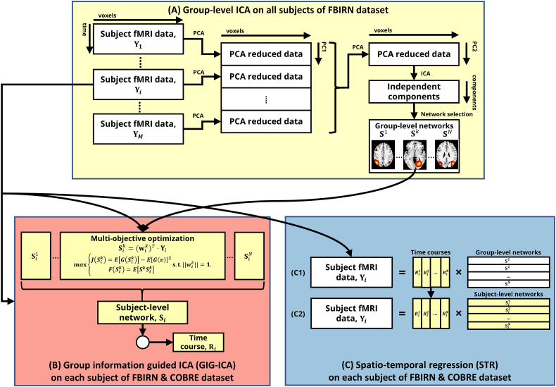
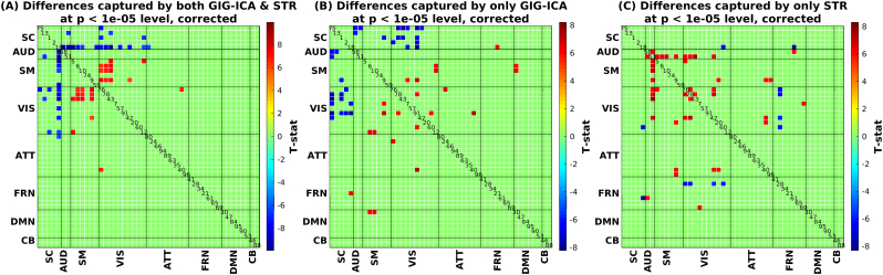
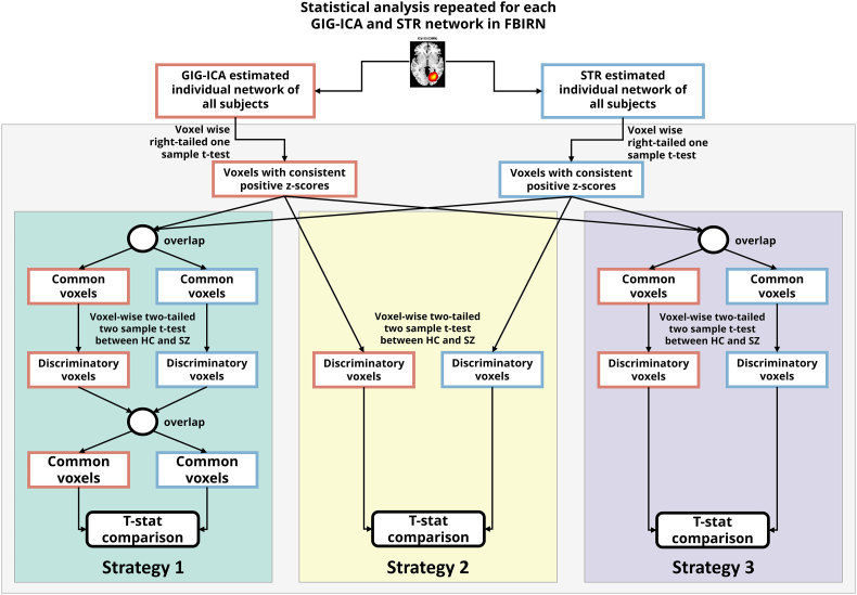
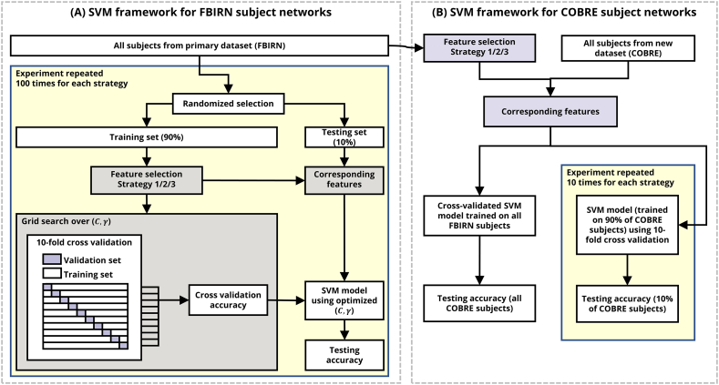
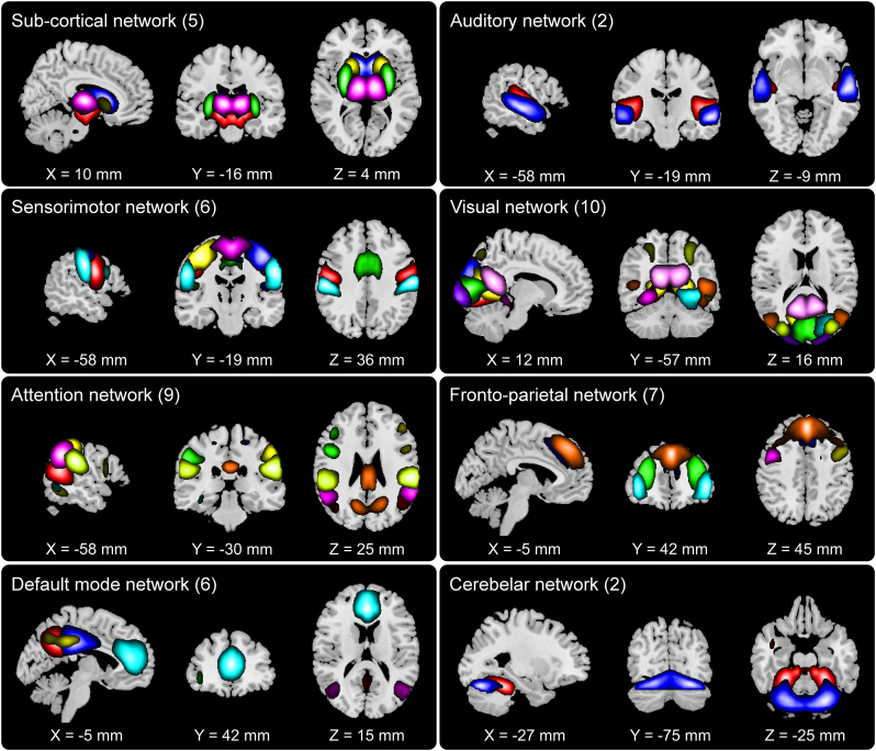
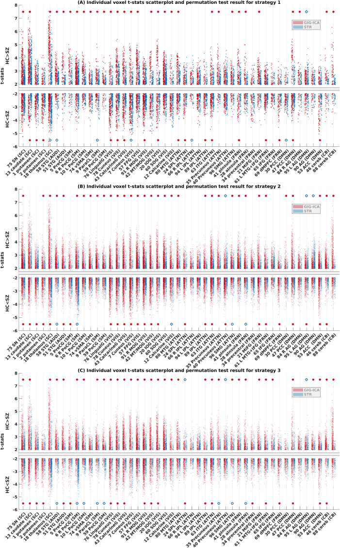
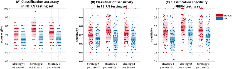
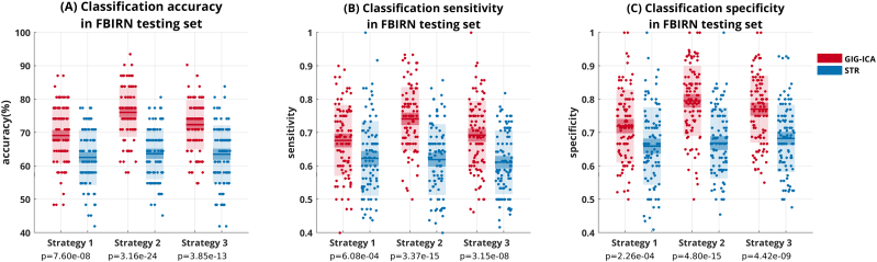
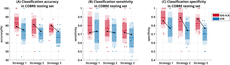
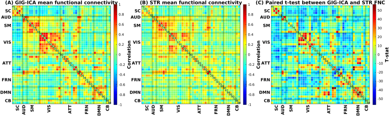
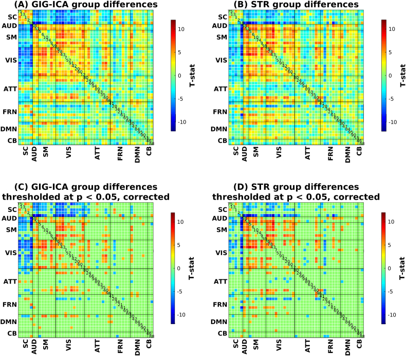
Brain functional networks identified from fMRI data can provide potential biomarkers for brain disorders. Group independent component analysis (GICA) is popular for extracting brain functional networks from multiple subjects. In GICA, different strategies exist for reconstructing subject-specific networks from the group-level networks. However, it is unknown whether these strategies have different sensitivities to group differences and abilities in distinguishing patients. Among GICA, spatio-temporal regression (STR) and spatially constrained ICA approaches such as group information guided ICA (GIG-ICA) can be used to propagate components (indicating networks) to a new subject that is not included in the original subjects. In this study, based on the same a priori network maps, we reconstructed subject-specific networks using these two methods separately from resting-state fMRI data of 151 schizophrenia patients (SZs) and 163 healthy controls (HCs). We investigated group differences in the estimated functional networks and the functional network connectivity (FNC) obtained by each method. The networks were also used as features in a cross-validated support vector machine (SVM) for classifying SZs and HCs. We selected features using different strategies to provide a comprehensive comparison between the two methods. GIG-ICA generally showed greater sensitivity in statistical analysis and better classification performance (accuracy 76.45 ± 8.9%, sensitivity 0.74 ± 0.11, specificity 0.79 ± 0.11) than STR (accuracy 67.45 ± 8.13%, sensitivity 0.65 ± 0.11, specificity 0.71 ± 0.11). Importantly, results were also consistent when applied to an independent dataset including 82 HCs and 82 SZs. Our work suggests that the functional networks estimated by GIG-ICA are more sensitive to group differences, and GIG-ICA is promising for identifying image-derived biomarkers of brain disease.
从 fMRI 数据中识别的脑功能网络可以为脑疾病提供潜在的生物标志物。组独立成分分析(GICA)是从多个被试中提取脑功能网络的常用方法。在 GICA 中,从组水平网络重建特定于个体的网络存在不同的策略。然而,尚不清楚这些策略对组间差异和区分患者的能力是否具有不同的敏感性。在 GICA 中,可以使用时空回归(STR)和空间约束 ICA 方法(如基于组信息的 ICA(GIG-ICA))将组件(表示网络)传播到原始被试中未包含的新被试。在这项研究中,基于相同的先验网络图谱,我们分别使用这两种方法从 151 名精神分裂症患者(SZ)和 163 名健康对照(HC)的静息态 fMRI 数据中重建特定于个体的网络。我们研究了估计的功能网络和每种方法获得的功能网络连接(FNC)中的组间差异。这些网络也被用作交叉验证支持向量机(SVM)的特征,用于分类 SZ 和 HC。我们使用不同的策略选择特征,以提供两种方法之间的全面比较。GIG-ICA 通常在统计分析中表现出更高的敏感性和更好的分类性能(准确率 76.45±8.9%,敏感性 0.74±0.11,特异性 0.79±0.11),优于 STR(准确率 67.45±8.13%,敏感性 0.65±0.11,特异性 0.71±0.11)。重要的是,当应用于包含 82 名 HC 和 82 名 SZ 的独立数据集时,结果也是一致的。我们的工作表明,GIG-ICA 估计的功能网络对组间差异更敏感,GIG-ICA 有望识别大脑疾病的图像衍生生物标志物。