Ura Daniel P, Karbowniczek Joanna E, Szewczyk Piotr K, Metwally Sara, Kopyściański Mateusz, Stachewicz Urszula
International Centre of Electron Microscopy for Materials Science, Faculty of Metals Engineering and Industrial Computer Science, AGH University of Science and Technology, 30-059 Krakow, Poland.
Faculty of Metals Engineering and Industrial Computer Science, AGH University of Science and Technology, 30-059 Krakow, Poland.
Bioengineering (Basel). 2019 May 9;6(2):41. doi: 10.3390/bioengineering6020041.
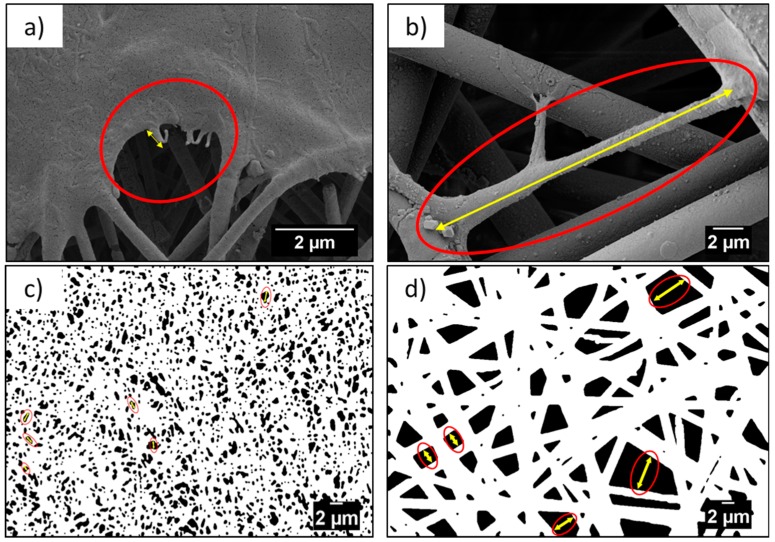
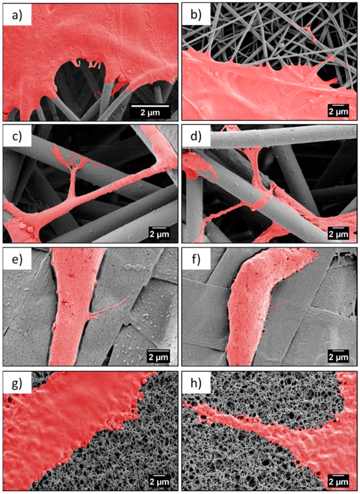
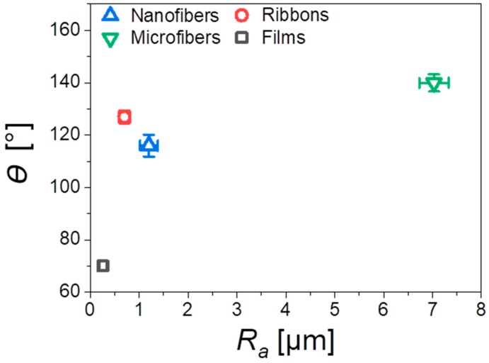
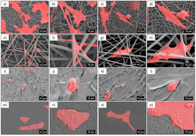
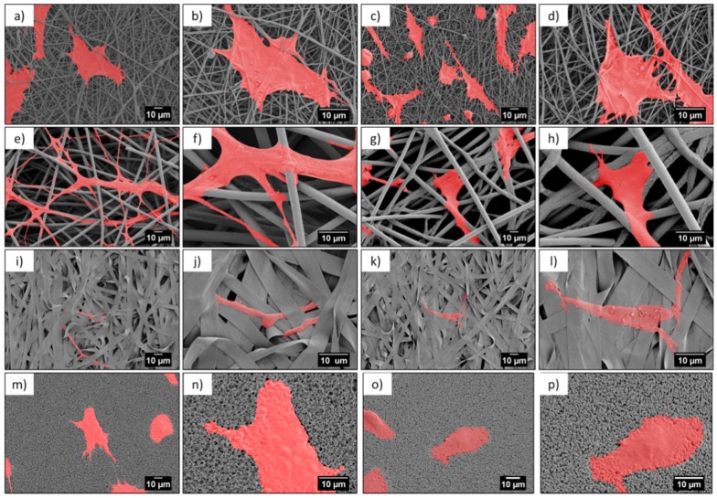
Tissue engineering requires properly selected geometry and surface properties of the scaffold, to promote in vitro tissue growth. In this study, we obtained three types of electrospun poly(methyl methacrylate) (PMMA) scaffolds-nanofibers, microfibers, and ribbons, as well as spin-coated films. Their morphology was imaged by scanning electron microscopy (SEM) and characterized by average surface roughness and water contact angle. PMMA films had a smooth surface with roughness, below 0.3 µm and hydrophilic properties, whereas for the fibers and the ribbons, we observed increased hydrophobicity, with higher surface roughness and fiber diameter. For microfibers, we obtained the highest roughness of 7 µm, therefore, the contact angle was 140°. All PMMA samples were used for the in vitro cell culture study, to verify the cells integration with various designs of scaffolds. The detailed microscopy study revealed that higher surface roughness enhanced cells' attachment and their filopodia length. The 3D structure of PMMA microfibers with an average fiber diameter above 3.5 µm, exhibited the most favorable geometry for cells' ingrowth, whereas, for other structures we observed cells growth only on the surface. The study showed that electrospinning of various scaffolds geometry is able to control cells development that can be adjusted according to the tissue needs in the regeneration processes.
组织工程需要对支架的几何形状和表面特性进行适当选择,以促进体外组织生长。在本研究中,我们获得了三种类型的电纺聚甲基丙烯酸甲酯(PMMA)支架——纳米纤维、微纤维和带状物,以及旋涂膜。通过扫描电子显微镜(SEM)对它们的形态进行成像,并通过平均表面粗糙度和水接触角进行表征。PMMA膜具有光滑表面,粗糙度低于0.3 µm且具有亲水性,而对于纤维和带状物,我们观察到疏水性增加,表面粗糙度和纤维直径更高。对于微纤维,我们获得了7 µm的最高粗糙度,因此接触角为140°。所有PMMA样品都用于体外细胞培养研究,以验证细胞与各种设计的支架的整合情况。详细的显微镜研究表明,较高的表面粗糙度增强了细胞的附着及其丝状伪足的长度。平均纤维直径大于3.5 µm的PMMA微纤维的三维结构,表现出对细胞向内生长最有利的几何形状,而对于其他结构,我们仅观察到细胞在表面生长。该研究表明,电纺各种支架几何形状能够控制细胞发育,可根据再生过程中的组织需求进行调整。