Vidavsky Netta, Kunitake Jennie A M R, Diaz-Rubio Maria Elena, Chiou Aaron E, Loh Hyun-Chae, Zhang Sheng, Masic Admir, Fischbach Claudia, Estroff Lara A
Department of Materials Science and Engineering, Cornell University, Ithaca, New York 14850, United States.
Metabolomics Facility, Institute of Biotechnology, Cornell University, Ithaca, New York 14850, United States.
ACS Cent Sci. 2019 May 22;5(5):768-780. doi: 10.1021/acscentsci.8b00932. Epub 2019 Apr 19.
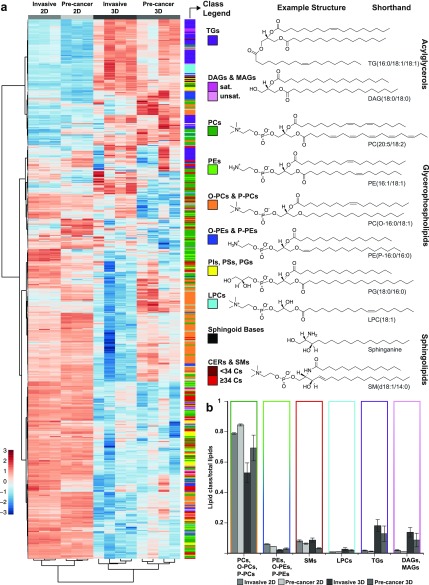
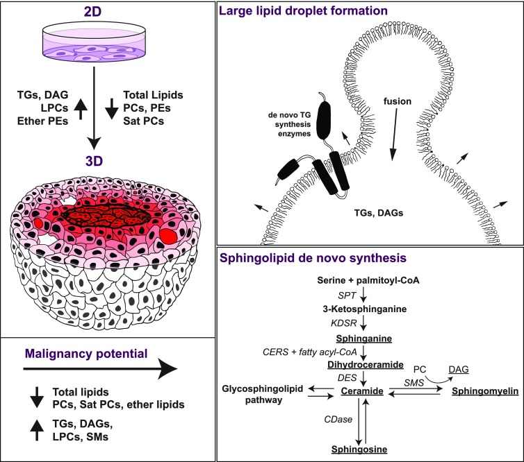
Aberrant lipid accumulation and marked changes in cellular lipid profiles are related to breast cancer metabolism and disease progression. , these phenomena are primarily studied using cells cultured in monolayers (2D). Here, we employ multicellular spheroids, generated using the MCF10A cell line series of increasing malignancy potential, to better recapitulate the 3D microenvironmental conditions that cells experience . Breast cancer cell lipid compositions were assessed in 2D and 3D culture models as a function of malignancy using liquid chromatography coupled with mass spectrometry. Further, the spatial distribution of lipids was examined using Raman chemical imaging and lipid staining. We show that with changes in the cellular microenvironment when moving from 2D to 3D cell cultures, total lipid amounts decrease significantly, while the ratio of acylglycerols to membrane lipids increases. This ratio increase could be associated with the formation of large lipid droplets (>10 μm) that are spatially evident throughout the spheroids but absent in 2D cultures. Additionally, we found a significant difference in lipid profiles between the more and less malignant spheroids, including changes that support sphingolipid production and a reduction in ether-linked lipid fractions in the invasive spheroids. These differences in lipid profiles as a function of cell malignancy and microenvironment highlight the importance of coupled spatial and lipidomic studies to better understand the connections between lipid metabolism and cancer.
异常的脂质积累和细胞脂质谱的显著变化与乳腺癌的代谢及疾病进展相关。目前,这些现象主要是利用单层培养的细胞(二维培养)进行研究。在此,我们采用由恶性潜能递增的MCF10A细胞系生成的多细胞球体,以更好地模拟细胞所经历的三维微环境条件。使用液相色谱-质谱联用技术,在二维和三维培养模型中评估乳腺癌细胞脂质组成与恶性程度的关系。此外,利用拉曼化学成像和脂质染色检查脂质的空间分布。我们发现,从二维细胞培养转变为三维细胞培养时,随着细胞微环境的变化,总脂质含量显著降低,而酰基甘油与膜脂的比例增加。这一比例的增加可能与大脂质滴(>10μm)的形成有关,这些大脂质滴在整个球体中在空间上明显可见,但在二维培养中不存在。此外,我们发现恶性程度较高和较低的球体之间脂质谱存在显著差异,包括支持鞘脂生成的变化以及侵袭性球体中醚键连接脂质组分的减少。这些脂质谱随细胞恶性程度和微环境的变化突出了结合空间和脂质组学研究以更好地理解脂质代谢与癌症之间联系的重要性。