Institute of Photonics and Photon-Technology, Northwest University, Xi'an 710069, China.
Department of Physics, Northwest University, Xi'an 710069, China.
Molecules. 2019 May 30;24(11):2059. doi: 10.3390/molecules24112059.
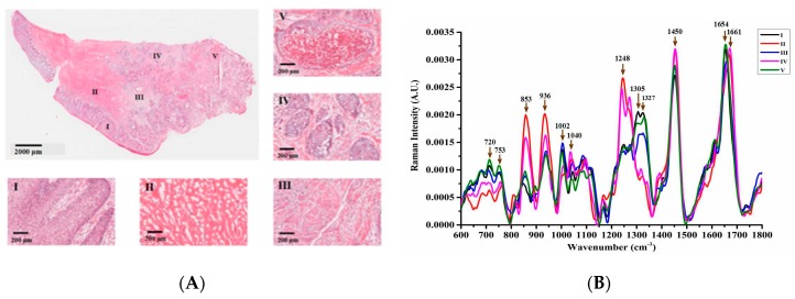
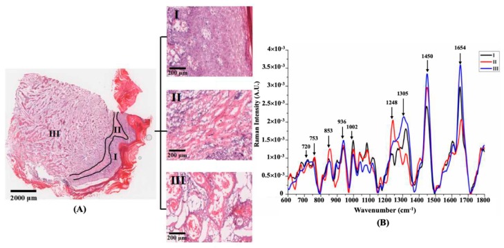
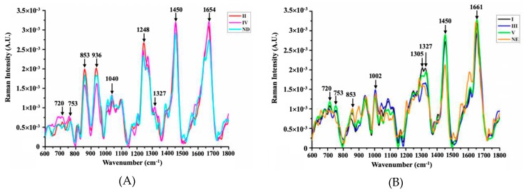
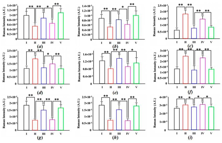
Raman spectroscopy facilitates accurate and minimally invasive investigation on biomedical samples to reveal their molecular-level biological information. In this work, the cancer field effects of squamous cell carcinoma (SCC) tissues were illustrated by Raman microspectroscopy. Referenced with hematoxylin and eosin (H&E) stained microscopic images, the biochemical variations during SCC progress were meticulously described by the Raman spectral features in different pathological areas of two lesion types, including the biochemical changes in collagen, lipids, DNA, and other components of SCC diffusion and metastasis. The experimental results demonstrated that the intensities of the Raman peaks representing collagen (853, 936, and 1248 cm) were decreased, whereas the intensities of peaks corresponding to DNA (720, 1327 cm) and lipids (1305 cm) were increased significantly in cancerous lesions, which testified that SCC originates from the epidermis and invades the dermis gradually. The achieved results not only described the molecular mechanism of skin carcinogenesis, but also provided vital reference data for in vivo skin cancer diagnosis using Raman spectroscopy.
拉曼光谱技术促进了对生物医学样本的准确和微创研究,以揭示其分子水平的生物信息。在这项工作中,通过拉曼显微光谱技术说明了鳞状细胞癌(SCC)组织的癌症领域效应。参考苏木精和伊红(H&E)染色的显微镜图像,通过两种病变类型不同病理区域的拉曼光谱特征详细描述了 SCC 进展过程中的生化变化,包括 SCC 扩散和转移中胶原蛋白、脂质、DNA 和其他成分的生化变化。实验结果表明,代表胶原蛋白的拉曼峰强度(853、936 和 1248 cm)降低,而对应于 DNA(720、1327 cm)和脂质(1305 cm)的峰强度显著增加,这表明 SCC 起源于表皮并逐渐侵袭真皮。所获得的结果不仅描述了皮肤癌变的分子机制,而且为使用拉曼光谱进行体内皮肤癌诊断提供了重要的参考数据。