Čepa Martin
Contipro a.s., Dolní Dobrouč 401, Dolní Dobrouč 561 02, Czech Republic.
Methods Protoc. 2018 Nov 19;1(4):43. doi: 10.3390/mps1040043.
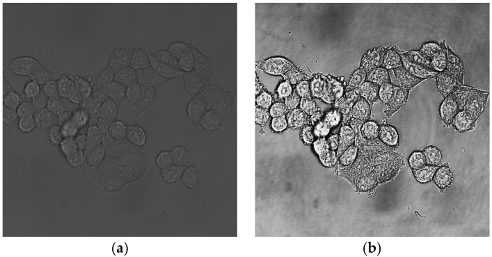
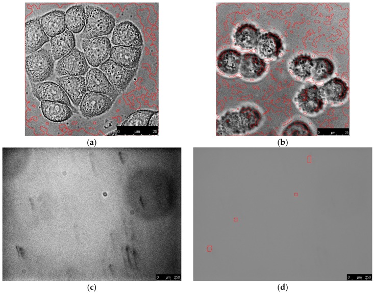
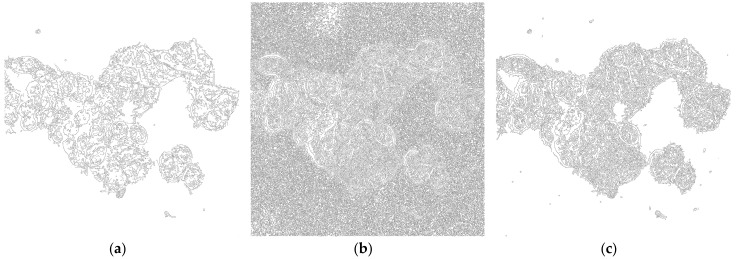
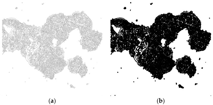
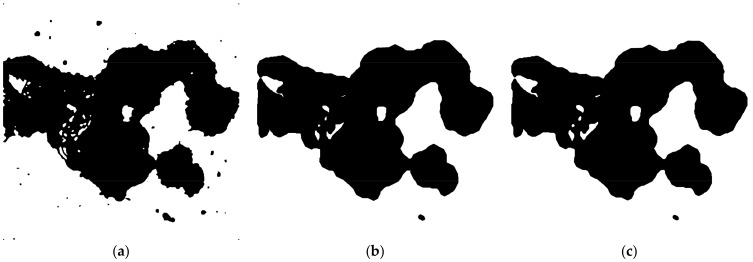
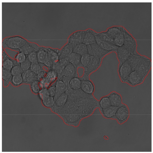
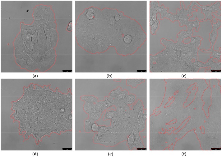
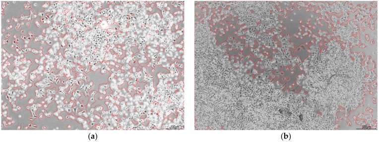
Segmentation is one of the most important steps in microscopy image analysis. Unfortunately, most of the methods use fluorescence images for this task, which is not suitable for analysis that requires a knowledge of area occupied by cells and an experimental design that does not allow necessary labeling. In this protocol, we present a simple method, based on edge detection and morphological operations, that separates total area occupied by cells from the background using only brightfield channel image. The resulting segmented picture can be further used as a mask for fluorescence quantification and other analyses. The whole procedure is carried out in open source software Fiji.
分割是显微镜图像分析中最重要的步骤之一。不幸的是,大多数方法都使用荧光图像来完成此任务,这不适用于需要了解细胞所占面积且实验设计不允许进行必要标记的分析。在本方案中,我们提出了一种基于边缘检测和形态学操作的简单方法,该方法仅使用明场通道图像就能将细胞所占的总面积与背景分离。所得的分割图像可进一步用作荧光定量和其他分析的掩膜。整个过程在开源软件Fiji中进行。