Lin Tai-Chi, Wang Lei-Chi, Yue Lan, Zhang Yi, Falabella Paulo, Zhu Danhong, Hinton David R, Rao Narsing A, Birch David G, Spencer Rand, Dorn Jessy D, Humayun Mark S
Department of Ophthalmology, USC Roski Eye Institute, University of Southern California, Los Angeles, CA, USA.
USC Ginsburg Institute for Biomedical Therapeutics, University of Southern California, Los Angeles, CA, USA.
Transl Vis Sci Technol. 2019 May 30;8(3):31. doi: 10.1167/tvst.8.3.31. eCollection 2019 May.
To characterize histologic changes in the optic nerve and the retina of an end-stage retinitis pigmentosa (RP) patient after long-term implantation with the Argus II retinal prosthesis system.
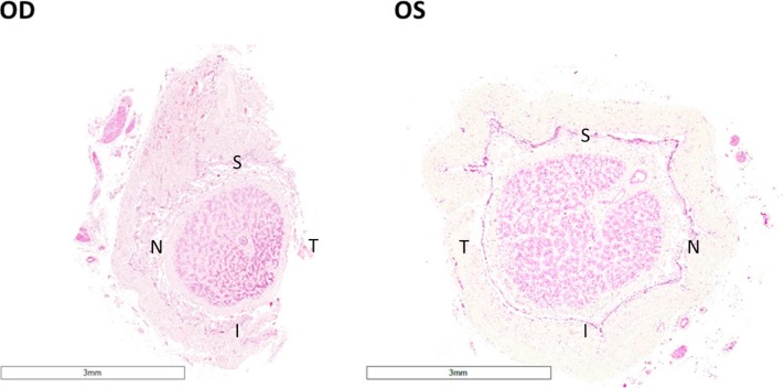
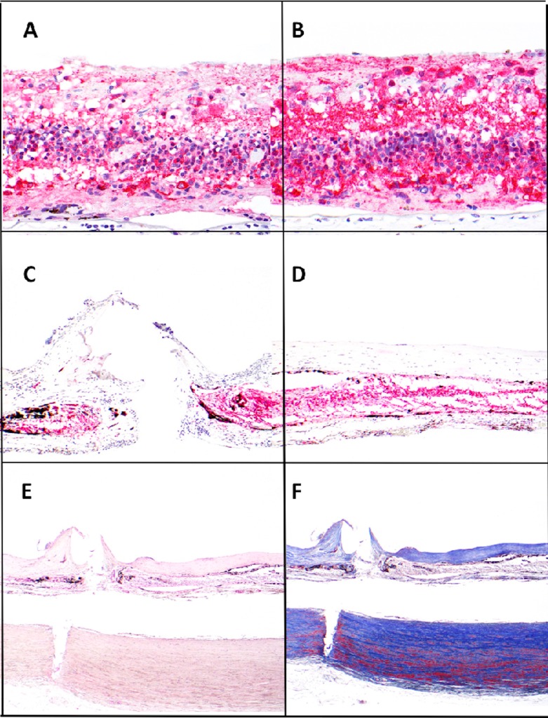
Serial cross sections from the patient's both eyes were collected postmortem 6 years after implantation. Optic nerve from both eyes were morphometrically analyzed and compared. Retina underneath and outside the array was analyzed and compared with corresponding regions in the fellow eye.
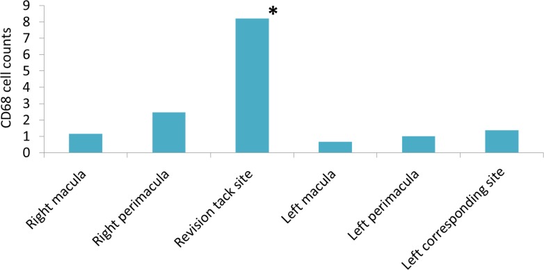
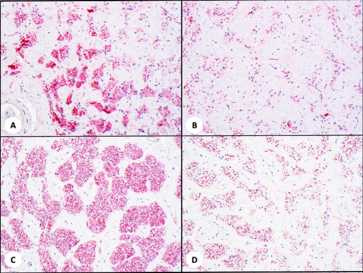
Although the optic nerve of the implant eye demonstrated significantly more overall atrophy than the fellow eye ( < 0.01), the temporal quadrant that retinotopically corresponded to the location of the array did not show additional damage. The total neuron count of the macular area was not significantly different between the two eyes, but the tack locations and their adjacent areas showed significantly fewer neurons than other perimacular areas. There was an increased expression of glial fibrillary acidic protein (GFAP) throughout the retina in the implant eye versus the fellow eye, but there was no significant difference in the cellular retinaldehyde-binding protein (CRALBP) expression. Except for the revision tack site, no significant increase of inflammatory reaction was detected in the implant eye.
Long-term implantation and electrical stimulation with an Argus II retinal prosthesis system did not result in significant tissue damage that could be detected by a morphometric analysis.
This study supports the long-term safety of the Argus II device and encourages further development of bioelectronics devices at the retina-machine interface.
描述长期植入阿格斯II型视网膜假体系统后,一名晚期视网膜色素变性(RP)患者视神经和视网膜的组织学变化。
在植入6年后对患者双眼进行尸检,收集连续的横断面切片。对双眼视神经进行形态计量分析并比较。分析植入阵列下方和外侧的视网膜,并与对侧眼的相应区域进行比较。
尽管植入眼的视神经整体萎缩程度明显高于对侧眼(<0.01),但在视网膜拓扑学上与阵列位置相对应的颞侧象限未显示出额外损伤。双眼黄斑区的神经元总数无显著差异,但固定钉位置及其相邻区域的神经元明显少于其他黄斑周边区域。与对侧眼相比,植入眼整个视网膜中胶质纤维酸性蛋白(GFAP)的表达增加,但细胞视黄醛结合蛋白(CRALBP)的表达无显著差异。除了修复固定钉部位外,在植入眼中未检测到炎症反应显著增加。
长期植入阿格斯II型视网膜假体系统并进行电刺激,未导致形态计量分析可检测到的显著组织损伤。
本研究支持阿格斯II型装置的长期安全性,并鼓励在视网膜-机器界面进一步开发生物电子装置。