Goudie Christine, Kinnin Jason, Bartellas Michael, Gullipalli Ravindra, Dubrowski Adam
Medical Education and Simulation, Memorial University of Newfoundland, St. John's, CAN.
Radiology, University of Saskatchewan College of Medicine, Saskatoon, USA.
Cureus. 2019 Apr 3;11(4):e4381. doi: 10.7759/cureus.4381.
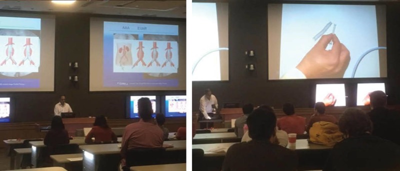
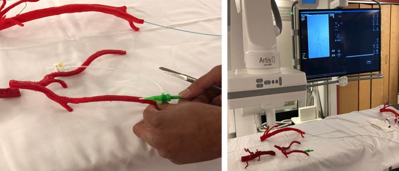
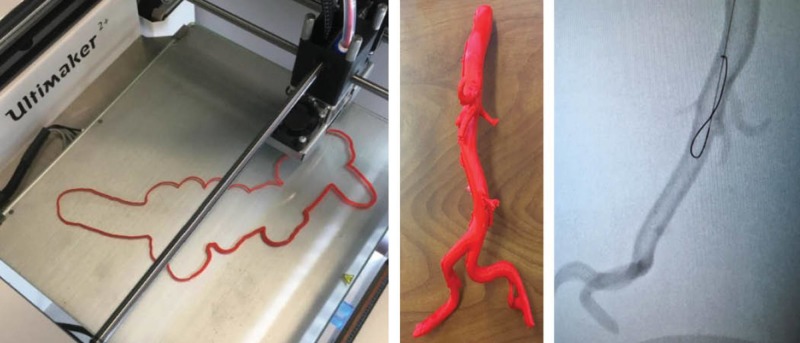
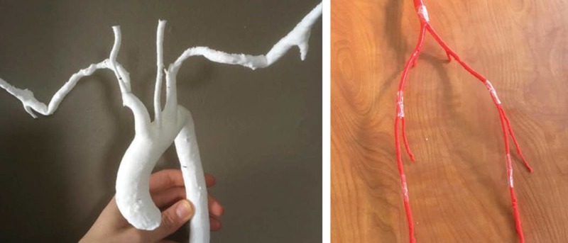
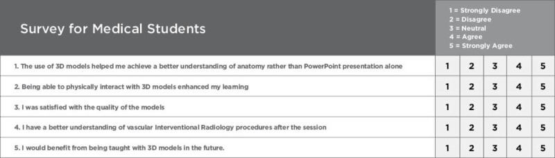
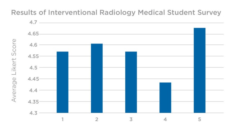
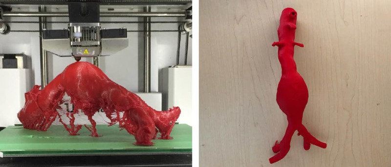
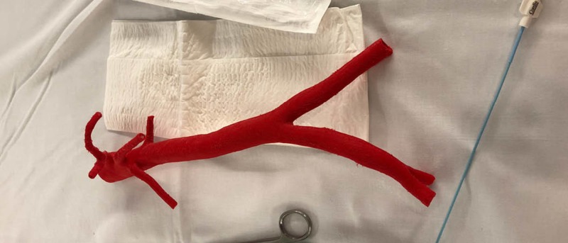
Three-dimensional (3D) printing has become a useful tool within the field of medicine as a way to produce custom anatomical models for teaching, surgical planning, and patient education. This technology is quickly becoming a key component in simulation-based medical education (SBME) to teach hands-on spatial perception and tactile feedback. Within fields such as interventional radiology (IR), this approach to SBME is also thought to be an ideal instructional method, providing an accurate and economical means to study human anatomy and vasculature. Such anatomical details can be extracted from patient-specific and anonymized CT or MRI scans for the purpose of teaching or analyzing patient-specific anatomy. There is evidence that 3D printing in IR can also optimize procedural training, so learners can rehearse procedures under fluoroscopy while receiving immediate supervisory feedback. Such training advancements in IR hold the potential to reduce procedural operating time, thus reducing the amount of time a patient is exposed to radiation and anaesthetia. Using a program evaluation approach, the purpose of this technical report is to describe the development and application of 3D-printed vasculature models within a radiology interest group to determine their efficacy as supplementary learning tools to traditional, lecture-based teaching. The study involved 30 medical students of varying years in their education, involved in the interest group at Memorial University of Newfoundland (MUN). The session was one hour in length and began with a Powerpoint presentation demonstrating the insertion of guide wires and stents using 3D-printed vasculature models. Participants had the opportunity to use the models to attempt several procedures demonstrated during the lecture. These attempts were supervised by an educational expert/facilitator. A survey was completed by all 30 undergraduate medical students and returned to the facilitators, who compiled the quantitative data to evaluate the efficacy of the 3D-printed models as an adjunct to the traditional didactic teaching within IR. The majority of feedback was positive, supporting the use of 3D=printed vasculature as an additional tactile training method for medical students within an IR academic setting. The hands-on experience provides a valuable training approach, with more opportunities for the rehearsal of high-acuity, low-occurrence (HALO) procedures performed in IR.
三维(3D)打印已成为医学领域的一种有用工具,可用于制作定制的解剖模型,用于教学、手术规划和患者教育。这项技术正迅速成为基于模拟的医学教育(SBME)中的关键组成部分,用于教授实践空间感知和触觉反馈。在介入放射学(IR)等领域,这种SBME方法也被认为是一种理想的教学方法,为研究人体解剖结构和脉管系统提供了一种准确且经济的手段。此类解剖细节可从患者特定的匿名CT或MRI扫描中提取,用于教学或分析患者特定的解剖结构。有证据表明,IR中的3D打印还可以优化程序训练,这样学习者就可以在荧光透视下演练程序,同时获得即时的监督反馈。IR中的此类训练进展有可能减少手术操作时间,从而减少患者接受辐射和麻醉的时间。本技术报告采用项目评估方法,旨在描述放射学兴趣小组内3D打印脉管系统模型的开发与应用,以确定其作为传统讲座式教学辅助学习工具的有效性。该研究涉及30名不同年级的医学生,他们都参与了纽芬兰纪念大学(MUN)的兴趣小组。课程时长为一小时,开始时是一个PowerPoint演示,展示了如何使用3D打印脉管系统模型插入导丝和支架。参与者有机会使用这些模型尝试讲座中演示的几种操作。这些尝试由一位教育专家/指导者进行监督。所有30名本科医学生都完成了一项调查,并将结果反馈给指导者,指导者汇总定量数据,以评估3D打印模型作为IR中传统理论教学辅助工具的有效性。大多数反馈都是积极的,支持将3D打印脉管系统作为IR学术环境中医学生的一种额外触觉训练方法。这种实践经验提供了一种有价值的训练方法,为在IR中进行的高敏锐度、低发生率(HALO)操作的演练提供了更多机会。