Fragogeorgi Eirini A, Rouchota Maritina, Georgiou Maria, Velez Marisela, Bouziotis Penelope, Loudos George
Institute of Nuclear & Radiological Sciences and Technology, Energy & Safety (INRASTES), NCSR "Demokritos", Athens, Greece.
Bioemission Technology Solutions (BIOEMTECH), Athens, Greece / Lefkippos Attica Technology Park, NCSR "Demokritos", Athens, Greece.
J Tissue Eng. 2019 Jun 21;10:2041731419854586. doi: 10.1177/2041731419854586. eCollection 2019 Jan-Dec.
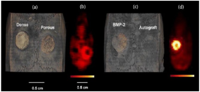
Bone is a dynamic tissue that constantly undergoes modeling and remodeling. Bone tissue engineering relying on the development of novel implant scaffolds for the treatment of pre-clinical bone defects has been extensively evaluated by histological techniques. The study of bone remodeling, that takes place over several weeks, is limited by the requirement of a large number of animals and time-consuming and labor-intensive procedures. X-ray-based imaging methods that can non-invasively detect the newly formed bone tissue have therefore been extensively applied in pre-clinical research and in clinical practice. The use of other imaging techniques at a pre-clinical level that act as supportive tools is convenient. This review mainly focuses on nuclear imaging methods (single photon emission computed tomography and positron emission tomography), either alone or used in combination with computed tomography. It addresses their application to small animal models with bone defects, both untreated and filled with substitute materials, to boost the knowledge on bone regenerative processes.
骨骼是一种动态组织,不断进行塑形和重塑。依靠新型植入支架的开发来治疗临床前骨缺损的骨组织工程已通过组织学技术进行了广泛评估。发生在数周内的骨重塑研究受到大量动物需求以及耗时且劳动密集型程序的限制。因此,能够非侵入性检测新形成骨组织的基于X射线的成像方法已广泛应用于临床前研究和临床实践。在临床前水平使用其他作为辅助工具的成像技术很方便。本综述主要关注核成像方法(单光子发射计算机断层扫描和正电子发射断层扫描),单独使用或与计算机断层扫描结合使用。它探讨了这些方法在有骨缺损的小动物模型中的应用,这些模型既未治疗也填充了替代材料,以增进对骨再生过程的了解。