• 文献检索
  • 文档翻译
  • 深度研究
  • 学术资讯
  • Suppr Zotero 插件Zotero 插件
  • 邀请有礼
  • 套餐&价格
  • 历史记录
应用&插件
Suppr Zotero 插件Zotero 插件浏览器插件Mac 客户端Windows 客户端微信小程序
定价
高级版会员购买积分包购买API积分包
服务
文献检索文档翻译深度研究API 文档MCP 服务
关于我们
关于 Suppr公司介绍联系我们用户协议隐私条款
关注我们

Suppr 超能文献

核心技术专利:CN118964589B侵权必究
粤ICP备2023148730 号-1Suppr @ 2026

文献检索

告别复杂PubMed语法,用中文像聊天一样搜索,搜遍4000万医学文献。AI智能推荐,让科研检索更轻松。

立即免费搜索

文件翻译

保留排版,准确专业,支持PDF/Word/PPT等文件格式,支持 12+语言互译。

免费翻译文档

深度研究

AI帮你快速写综述,25分钟生成高质量综述,智能提取关键信息,辅助科研写作。

立即免费体验

通过软骨内成骨治疗节段性骨缺损的疗效:3D打印设计与骨代谢活动

Efficacy of treating segmental bone defects through endochondral ossification: 3D printed designs and bone metabolic activities.

作者信息

Hara Kenji, Hellem Endre, Yamada Shuntaro, Sariibrahimoglu Kemal, Mølster Anders, Gjerdet Nils R, Hellem Sølve, Mustafa Kamal, Yassin Mohammed A

机构信息

Centre of Translational Oral Research (TOR) - Tissue Engineering Group, Department of Clinical Dentistry, University of Bergen, Bergen, Norway.

Department of Oral and Maxillofacial Surgery, Fujieda Heisei Memorial Hospital, Japan.

出版信息

Mater Today Bio. 2022 Mar 7;14:100237. doi: 10.1016/j.mtbio.2022.100237. eCollection 2022 Mar.

DOI:10.1016/j.mtbio.2022.100237
PMID:35280332
原文链接:https://pmc.ncbi.nlm.nih.gov/articles/PMC8914554/
Abstract

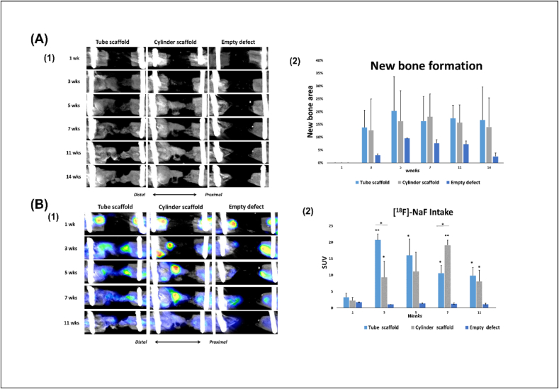
Three-dimensional printing (3D printing) is a promising technique for producing scaffolds for bone tissue engineering applications. Porous scaffolds can be printed directly, and the design, shape and porosity can be controlled. 3D synthetic biodegradable polymeric scaffolds intended for in situ bone regeneration must meet stringent criteria, primarily appropriate mechanical properties, good 3D design, adequate biocompatibility and the ability to enhance bone formation. In this study, healing of critical-sized (5 ​mm) femur defects of rats was enhanced by implanting two different designs of 3D printed poly(l-lactide-co-ε-caprolactone) (poly(LA-co-CL)) scaffolds seeded with rat bone marrow mesenchymal stem cells (rBMSC), which had been pre-differentiated into cartilage-forming chondrocytes. Depending on the design, the scaffolds had an interconnected porous structure of 300-500 ​μm and porosity of 50-65%. According to a computational simulation, the internal force distribution was consistent with scaffold designs and comparable between the two designs. Moreover, the defects treated with 3D-printed scaffolds seeded with chondrocyte-like cells exhibited significantly increased bone formation up to 15 weeks compared with empty defects. In all experimental animals, bone metabolic activity was monitored by positron emission tomography 1, 3, 5, 7, 11 and 14 weeks after surgery. This demonstrated a time-dependent relationship between scaffold design and metabolic activity. This confirmed that successful regeneration was highly reproducible. The and data indicated that the experimental setups had promising outcomes and could facilitate new bone formation through endochondral ossification.

摘要

三维打印(3D打印)是一种很有前景的技术,可用于制造用于骨组织工程应用的支架。多孔支架可以直接打印,其设计、形状和孔隙率均可控制。用于原位骨再生的3D合成可生物降解聚合物支架必须满足严格的标准,主要包括合适的机械性能、良好的三维设计、足够的生物相容性以及促进骨形成的能力。在本研究中,通过植入两种不同设计的3D打印聚(L-丙交酯-共-ε-己内酯)(聚(LA-co-CL))支架,增强了大鼠临界尺寸(5毫米)股骨缺损的愈合,这些支架接种了已预分化为软骨形成软骨细胞的大鼠骨髓间充质干细胞(rBMSC)。根据设计不同,支架具有300-500μm的相互连接的多孔结构,孔隙率为50-65%。根据计算模拟,内力分布与支架设计一致,两种设计之间具有可比性。此外,与空白缺损相比,用接种软骨样细胞的3D打印支架治疗的缺损在长达15周的时间里骨形成显著增加。在所有实验动物中,术后1、3、5、7、11和14周通过正电子发射断层扫描监测骨代谢活性。这证明了支架设计与代谢活性之间存在时间依赖性关系。这证实了成功的再生具有高度可重复性。 和 数据表明,实验设置具有良好的结果,并且可以通过软骨内骨化促进新骨形成。

https://cdn.ncbi.nlm.nih.gov/pmc/blobs/fff2/8914554/4864db67c8e2/gr7.jpg
https://cdn.ncbi.nlm.nih.gov/pmc/blobs/fff2/8914554/33398f83ffa1/ga1.jpg
https://cdn.ncbi.nlm.nih.gov/pmc/blobs/fff2/8914554/ae94200df6cf/gr1.jpg
https://cdn.ncbi.nlm.nih.gov/pmc/blobs/fff2/8914554/d16c5f679cda/gr2.jpg
https://cdn.ncbi.nlm.nih.gov/pmc/blobs/fff2/8914554/97c8eefc72d6/gr3.jpg
https://cdn.ncbi.nlm.nih.gov/pmc/blobs/fff2/8914554/390156ac1fd6/gr4.jpg
https://cdn.ncbi.nlm.nih.gov/pmc/blobs/fff2/8914554/0d19c58e8d97/gr5.jpg
https://cdn.ncbi.nlm.nih.gov/pmc/blobs/fff2/8914554/fc9f69ba2727/gr6.jpg
https://cdn.ncbi.nlm.nih.gov/pmc/blobs/fff2/8914554/4864db67c8e2/gr7.jpg

相似文献

1
Efficacy of treating segmental bone defects through endochondral ossification: 3D printed designs and bone metabolic activities.通过软骨内成骨治疗节段性骨缺损的疗效:3D打印设计与骨代谢活动
Mater Today Bio. 2022 Mar 7;14:100237. doi: 10.1016/j.mtbio.2022.100237. eCollection 2022 Mar.
2
Biodegradable poly(caprolactone fumarate) 3D printed scaffolds for segmental bone defects within the Masquelet technique.可生物降解的聚(富马酸己内酯)3D 打印支架用于 Masquelet 技术中的节段性骨缺损。
J Orthop Res. 2024 Sep;42(9):1974-1983. doi: 10.1002/jor.25839. Epub 2024 Mar 24.
3
Three-Dimensional Printing of Biodegradable Piperazine-Based Polyurethane-Urea Scaffolds with Enhanced Osteogenesis for Bone Regeneration.具有增强成骨作用的可生物降解哌嗪基聚氨酯脲支架的三维打印用于骨再生。
ACS Appl Mater Interfaces. 2019 Mar 6;11(9):9415-9424. doi: 10.1021/acsami.8b20323. Epub 2019 Feb 13.
4
Suppressing mesenchymal stem cell hypertrophy and endochondral ossification in 3D cartilage regeneration with nanofibrous poly(l-lactic acid) scaffold and matrilin-3.利用纳米纤维聚(L-乳酸)支架和软骨基质蛋白 3 抑制 3D 软骨再生中的间充质干细胞肥大和软骨内骨化。
Acta Biomater. 2018 Aug;76:29-38. doi: 10.1016/j.actbio.2018.06.027. Epub 2018 Jun 22.
5
3D Printed Chitosan Composite Scaffold for Chondrocytes Differentiation.3D 打印壳聚糖复合支架促进软骨细胞分化。
Curr Med Imaging. 2021;17(7):832-842. doi: 10.2174/1573405616666201217112939.
6
3D printing of metal-organic framework incorporated porous scaffolds to promote osteogenic differentiation and bone regeneration.用于促进成骨分化和骨再生的金属有机框架复合多孔支架的3D打印
Nanoscale. 2020 Dec 23;12(48):24437-24449. doi: 10.1039/d0nr06297a.
7
Dual-functional 3D-printed composite scaffold for inhibiting bacterial infection and promoting bone regeneration in infected bone defect models.用于抑制感染性骨缺损模型中的细菌感染和促进骨再生的双重功能 3D 打印复合支架。
Acta Biomater. 2018 Oct 1;79:265-275. doi: 10.1016/j.actbio.2018.08.015. Epub 2018 Aug 18.
8
3D-Printed composite scaffolds based on poly(ε-caprolactone) filled with poly(glutamic acid)-modified cellulose nanocrystals for improved bone tissue regeneration.基于聚(ε-己内酯)填充聚(谷氨酸)改性纤维素纳米晶体的 3D 打印复合支架用于改善骨组织再生。
J Biomed Mater Res B Appl Biomater. 2022 Nov;110(11):2422-2437. doi: 10.1002/jbm.b.35100. Epub 2022 May 26.
9
Wet 3D printing of biodegradable porous scaffolds to enable room-temperature deposition modeling of polymeric solutions for regeneration of articular cartilage.湿 3D 打印可生物降解多孔支架,实现室温下聚合溶液沉积成型,用于关节软骨再生。
Biofabrication. 2024 Apr 12;16(3). doi: 10.1088/1758-5090/ad3a12.
10
Low temperature hybrid 3D printing of hierarchically porous bone tissue engineering scaffolds withdelivery of osteogenic peptide and mesenchymal stem cells.低温杂化 3D 打印具有层级多孔结构的骨组织工程支架并递送成骨肽和间充质干细胞。
Biofabrication. 2022 Aug 11;14(4). doi: 10.1088/1758-5090/ac84b0.

引用本文的文献

1
Lactate-functionalized 3D-printed PCL/nHA scaffold drives BMSC osteogenesis via metabolic-epigenetic crosstalk.乳酸功能化3D打印PCL/nHA支架通过代谢-表观遗传串扰驱动骨髓间充质干细胞成骨分化。
Mater Today Bio. 2025 Jul 16;33:102101. doi: 10.1016/j.mtbio.2025.102101. eCollection 2025 Aug.
2
Exploring calcium-free alternatives in endochondral bone repair tested on trials - A review.探索用于软骨内骨修复的无钙替代物的试验研究综述
Regen Ther. 2024 Jun 1;26:145-160. doi: 10.1016/j.reth.2024.05.017. eCollection 2024 Jun.
3
Resistin targets TAZ to promote osteogenic differentiation through PI3K/AKT/mTOR pathway.

本文引用的文献

1
Induction of osteogenic differentiation of bone marrow stromal cells on 3D polyester-based scaffolds solely by subphysiological fluidic stimulation in a laminar flow bioreactor.在层流生物反应器中,仅通过亚生理流体刺激在基于三维聚酯的支架上诱导骨髓基质细胞的成骨分化。
J Tissue Eng. 2021 Jun 24;12:20417314211019375. doi: 10.1177/20417314211019375. eCollection 2021 Jan-Dec.
2
18F-NaF PET/CT imaging of bone formation induced by bioactive glass S53P4 after mastoid obliteration.乳突切除术后生物活性玻璃S53P4诱导骨形成的18F-NaF PET/CT成像
Eur J Hybrid Imaging. 2019 Oct 31;3(1):18. doi: 10.1186/s41824-019-0065-3.
3
抵抗素通过PI3K/AKT/mTOR信号通路作用于TAZ以促进成骨分化。
iScience. 2023 Jun 7;26(7):107025. doi: 10.1016/j.isci.2023.107025. eCollection 2023 Jul 21.
4
Regenerated silk fibroin based on small aperture scaffolds and marginal sealing hydrogel for osteochondral defect repair.基于小孔径支架和边缘封闭水凝胶的再生丝素蛋白用于骨软骨缺损修复。
Biomater Res. 2023 May 19;27(1):50. doi: 10.1186/s40824-023-00370-1.
5
Surgical Classification for Preclinical Rat Femoral Bone Defect Model: Standardization Based on Systematic Review, Anatomical Analysis and Virtual Surgery.临床前大鼠股骨骨缺损模型的手术分类:基于系统评价、解剖学分析和虚拟手术的标准化
Bioengineering (Basel). 2022 Sep 15;9(9):476. doi: 10.3390/bioengineering9090476.
6
3D-printed high-density polyethylene scaffolds with bioactive and antibacterial layer-by-layer modification for auricle reconstruction.用于耳廓重建的具有生物活性和抗菌逐层修饰的3D打印高密度聚乙烯支架
Mater Today Bio. 2022 Jul 15;16:100361. doi: 10.1016/j.mtbio.2022.100361. eCollection 2022 Dec.
Initial mechanical conditions within an optimized bone scaffold do not ensure bone regeneration - an in silico analysis.
优化的骨支架内的初始机械条件并不能确保骨再生——一种计算机模拟分析。
Biomech Model Mechanobiol. 2021 Oct;20(5):1723-1731. doi: 10.1007/s10237-021-01472-2. Epub 2021 Jun 7.
4
Additive Manufacturing of Material Scaffolds for Bone Regeneration: Toward Application in the Clinics.用于骨再生的材料支架的增材制造:迈向临床应用
Adv Funct Mater. 2020 Oct 15;31(5). doi: 10.1002/adfm.202006967. eCollection 2021 Jan 27.
5
Orthotopic Bone Regeneration within 3D Printed Bioceramic Scaffolds with Region-Dependent Porosity Gradients in an Equine Model.马模型中具有区域依赖性孔隙率梯度的3D打印生物陶瓷支架内的原位骨再生
Adv Healthc Mater. 2020 May;9(10):e1901807. doi: 10.1002/adhm.201901807. Epub 2020 Apr 23.
6
Biomaterial-based endochondral bone regeneration: a shift from traditional tissue engineering paradigms to developmentally inspired strategies.基于生物材料的软骨内骨再生:从传统组织工程范式向发育启发策略的转变。
Mater Today Bio. 2019 May 31;3:100009. doi: 10.1016/j.mtbio.2019.100009. eCollection 2019 Jun.
7
Printability and Critical Insight into Polymer Properties during Direct-Extrusion Based 3D Printing of Medical Grade Polylactide and Copolyesters.直接挤出式 3D 打印医用级聚乳酸及其共聚酯的可印刷性和对聚合物性能的关键洞察
Biomacromolecules. 2020 Feb 10;21(2):388-396. doi: 10.1021/acs.biomac.9b01112. Epub 2019 Oct 11.
8
Repair of Critical-Sized Long Bone Defects Using Dipyridamole-Augmented 3D-Printed Bioactive Ceramic Scaffolds.使用双嘧达莫增强的 3D 打印生物活性陶瓷支架修复临界尺寸长骨缺损。
J Orthop Res. 2019 Dec;37(12):2499-2507. doi: 10.1002/jor.24424. Epub 2019 Aug 8.
9
Robust bone regeneration through endochondral ossification of human mesenchymal stem cells within their own extracellular matrix.通过人骨髓间充质干细胞自身细胞外基质中的软骨内骨化实现健壮的骨再生。
Biomaterials. 2019 Oct;218:119336. doi: 10.1016/j.biomaterials.2019.119336. Epub 2019 Jul 6.
10
imaging techniques for bone tissue engineering.用于骨组织工程的成像技术。
J Tissue Eng. 2019 Jun 21;10:2041731419854586. doi: 10.1177/2041731419854586. eCollection 2019 Jan-Dec.