Department of Oral Medicine and Periodontology, Faculty of Dentistry, Mahidol University, Bangkok, 10400, Thailand.
Department of Oral and Maxillofacial Pathology, Faculty of Dentistry, Mahidol University, Bangkok, 10400, Thailand.
BMC Oral Health. 2019 Jul 10;19(1):142. doi: 10.1186/s12903-019-0832-3.
As oral cavity is the main location of Epstein-Barr virus (EBV) latency and shedding, and as EBV-encoded latent membrane protein-1 (LMP-1) has a crucial role in cell transformation, association between EBV infection, LMP-1 expression and oral malignancy is of interest. Although EBV DNA has been detected in oral squamous cell carcinoma (OSCC), studies on LMP-1 expression in OSCC and oral potentially malignant disorders are scarce and still controversial. This study aimed to evaluate the expression of LMP-1 in OSCC and oral leukoplakia (OL).
Biopsy specimens of 36 OSCC, 69 OL with and without dysplasia and 10 normal oral mucosa were assessed for the expression of LMP-1 using immunohistochemistry. In each case, at least 1000 cells were counted. Cells with staining were considered positive, classified by location as nuclear, cytoplasmic and nuclear plus cytoplasmic staining. Percentage of positive cells at different locations and of total positive cells were determined. For statistical analysis, SPSS version 21 was used. Statistical significance was considered at p < 0.05.
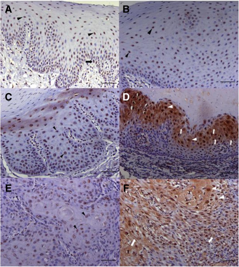
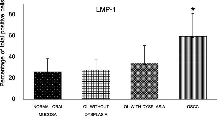
LMP-1 was expressed in all studied specimens. In terms of percentage of total positive cells, LMP-1 expression was higher from normal mucosa (26.36%), OL without dysplasia (28.03%), OL with dysplasia (34.15%), to the significantly highest, (59.67%) in OSCC. In addition, cells with nuclear staining alone, cytoplasmic staining alone and cells with nuclear plus cytoplasmic staining were significantly higher in OSCC compared to those of normal mucosa, OL with and without dysplasia.
LMP-1 was overexpressed in OSCC. Our analysis on subcellular localization of LMP-1 in OSCC revealed prominent distinguished pattern, cytoplasmic distribution. Further studies in cell lines and animals are required to clarify the association between this EBV-encoded proteins and oral carcinogenesis.
口腔是 EBV(Epstein-Barr 病毒)潜伏和脱落的主要部位,而 EBV 编码的潜伏膜蛋白 1(LMP-1)在细胞转化中起着关键作用,因此 EBV 感染、LMP-1 表达与口腔恶性肿瘤之间的关联备受关注。尽管已在口腔鳞状细胞癌(OSCC)中检测到 EBV DNA,但关于 OSCC 和口腔潜在恶性疾病中 LMP-1 表达的研究仍然很少,且存在争议。本研究旨在评估 LMP-1 在 OSCC 和口腔白斑(OL)中的表达。
使用免疫组织化学法检测 36 例 OSCC、69 例伴或不伴异型增生的 OL 和 10 例正常口腔黏膜的 LMP-1 表达。在每种情况下,至少计数 1000 个细胞。有染色的细胞被认为是阳性细胞,根据位置分为核、细胞质和核质共染。确定不同位置的阳性细胞百分比和总阳性细胞百分比。使用 SPSS 版本 21 进行统计分析。认为 p<0.05 具有统计学意义。
LMP-1 在所有研究标本中均有表达。就总阳性细胞的百分比而言,LMP-1 的表达从正常黏膜(26.36%)、无异型增生的 OL(28.03%)、有异型增生的 OL(34.15%)逐渐升高,到 OSCC 时达到最高水平(59.67%)。此外,与正常黏膜、无异型增生和有异型增生的 OL 相比,OSCC 中仅存在核染色、仅存在细胞质染色和核质共染的细胞比例明显更高。
LMP-1 在 OSCC 中过度表达。我们对 OSCC 中 LMP-1 亚细胞定位的分析显示出明显的细胞质分布模式。需要进一步在细胞系和动物中进行研究,以阐明这些 EBV 编码蛋白与口腔癌变之间的关联。