Aix Marseille Univ, CNRS, Centrale Marseille, Institut Fresnel, Marseille, France.
Institut Paoli-Calmettes, Endoscopy and Gastroenterology Departement, Marseille, France.
Sci Rep. 2019 Jul 11;9(1):10052. doi: 10.1038/s41598-019-46489-x.
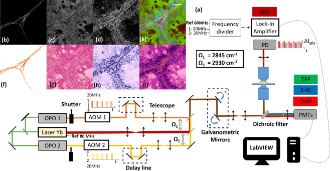
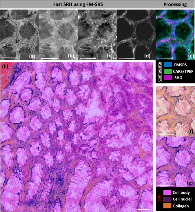
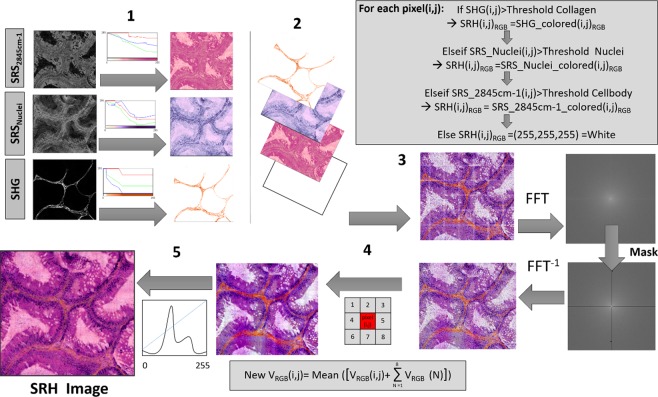
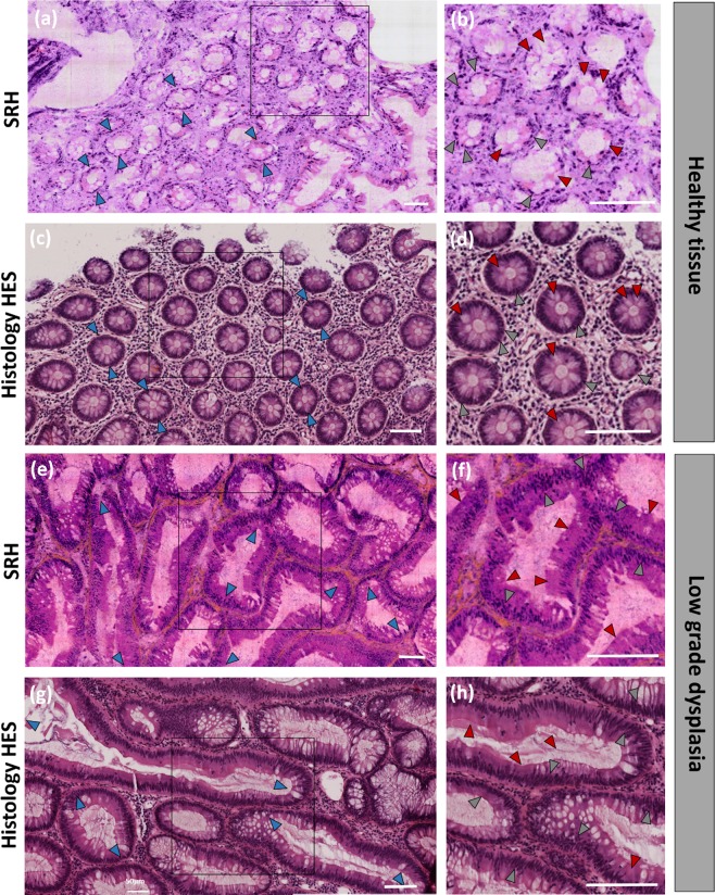
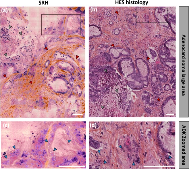
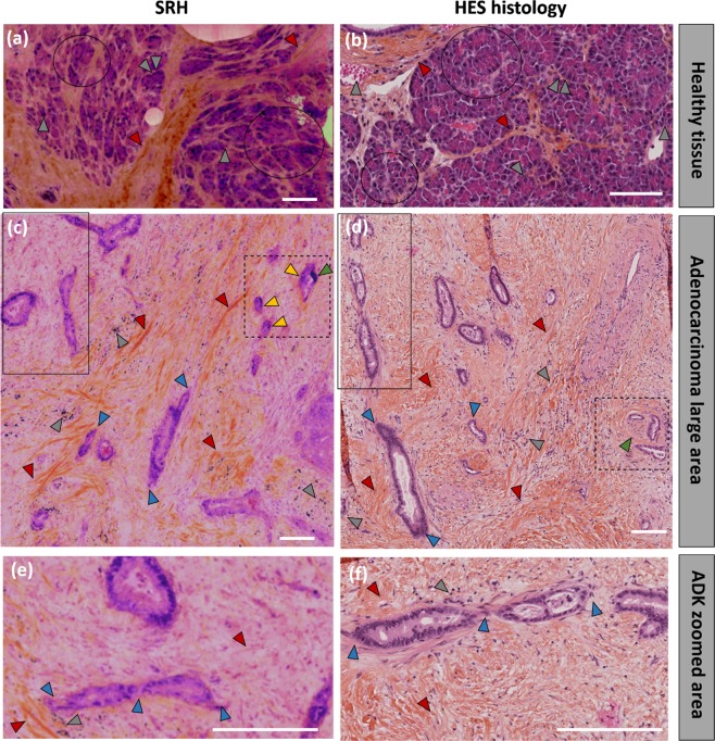
Conventional haematoxylin, eosin and saffron (HES) histopathology, currently the 'gold-standard' for pathological diagnosis of cancer, requires extensive sample preparations that are achieved within time scales that are not compatible with intra-operative situations where quick decisions must be taken. Providing to pathologists a close to real-time technology revealing tissue structures at the cellular level with HES histologic quality would provide an invaluable tool for surgery guidance with evident clinical benefit. Here, we specifically develop a stimulated Raman imaging based framework that demonstrates gastro-intestinal (GI) cancer detection of unprocessed human surgical specimens. The generated stimulated Raman histology (SRH) images combine chemical and collagen information to mimic conventional HES histopathology staining. We report excellent agreements between SRH and HES images acquire on the same patients for healthy, pre-cancerous and cancerous colon and pancreas tissue sections. We also develop a novel fast SRH imaging modality that captures at the pixel level all the information necessary to provide instantaneous SRH images. These developments pave the way for instantaneous label free GI histology in an intra-operative context.
常规苏木精-伊红-番红(HES)组织病理学目前是癌症病理诊断的“金标准”,需要进行广泛的样本制备,而这些制备过程在时间尺度上与术中情况不兼容,因为必须在术中快速做出决策。为病理学家提供一种接近实时的技术,以细胞水平的 HES 组织学质量揭示组织结构,将为手术指导提供一个非常有价值的工具,具有明显的临床益处。在这里,我们专门开发了一种基于受激发射拉曼成像的框架,该框架证明了对未经处理的人类手术标本进行胃肠(GI)癌症检测。生成的受激发射拉曼组织学(SRH)图像结合了化学和胶原信息,以模拟常规 HES 组织病理学染色。我们报告了在相同患者的健康、癌前和癌症结肠和胰腺组织切片上采集的 SRH 和 HES 图像之间的极好一致性。我们还开发了一种新的快速 SRH 成像方式,可以在像素级捕获提供即时 SRH 图像所需的所有信息。这些发展为术中即时进行无标记的 GI 组织学铺平了道路。