Lombardini Alberto, Mytskaniuk Vasyl, Sivankutty Siddharth, Andresen Esben Ravn, Chen Xueqin, Wenger Jérôme, Fabert Marc, Joly Nicolas, Louradour Frédéric, Kudlinski Alexandre, Rigneault Hervé
1Aix-Marseille Univ, CNRS, Centrale Marseille, Institut Fresnel, Marseille, France.
2Laboratoire de Physique des Lasers Atomes et Molécules, UMR 8523, CNRS, Université Lille, 59000 Lille, France.
Light Sci Appl. 2018 May 30;7:10. doi: 10.1038/s41377-018-0003-3. eCollection 2018.
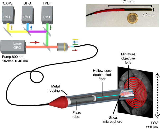
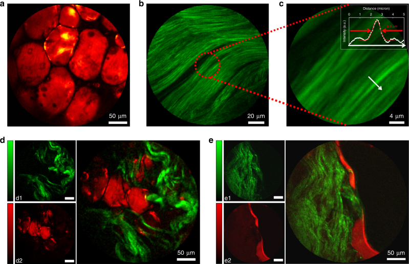
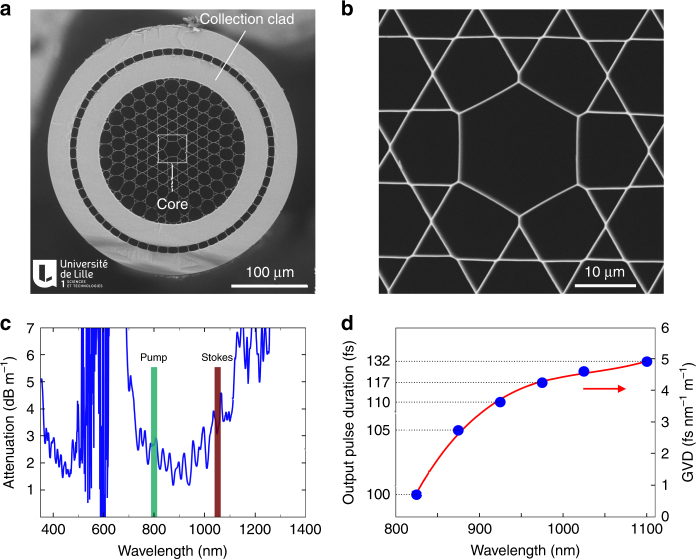
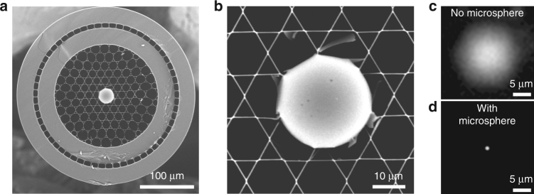
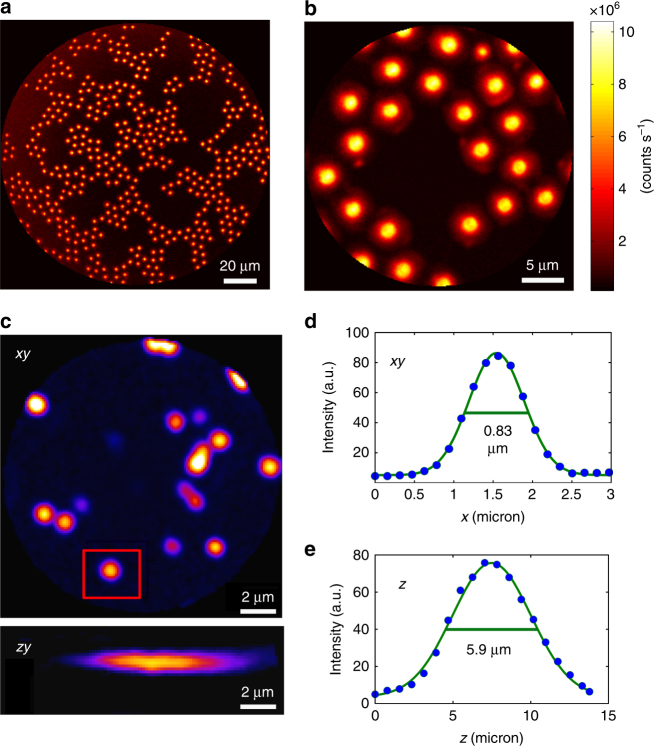
Coherent Raman scattering microscopy is a fast, label-free, and chemically specific imaging technique that shows high potential for future in vivo optical histology. However, the imaging depth in tissues is limited to the sub-millimeter range because of absorption and scattering. Realization of coherent Raman imaging using a fiber endoscope system is a crucial step towards imaging deep inside living tissues and providing information that is inaccessible with current microscopy tools. Until now, the development of coherent Raman endoscopy has been hampered by several issues, mainly related to the fiber delivery of the excitation pulses and signal collection. Here, we present a flexible, compact, coherent Raman, and multimodal nonlinear endoscope (4.2 mm outer diameter, 71 mm rigid length) based on a resonantly scanned hollow-core Kagomé-lattice double-clad fiber. The fiber design enables distortion-less, background-free delivery of femtosecond excitation pulses and back-collection of nonlinear signals through the same fiber. Sub-micrometer spatial resolution over a large field of view is obtained by combination of a miniature objective lens with a silica microsphere lens inserted into the fiber core. We demonstrate high-resolution, high-contrast coherent anti-Stokes Raman scattering, and second harmonic generation endoscopic imaging of biological tissues over a field of view of 320 µm at a rate of 0.8 frames per second. These results pave the way for intraoperative label-free imaging applied to real-time histopathology diagnosis and surgery guidance.
相干拉曼散射显微镜是一种快速、无标记且具有化学特异性的成像技术,在未来的体内光学组织学方面显示出巨大潜力。然而,由于吸收和散射,组织中的成像深度仅限于亚毫米范围。利用光纤内窥镜系统实现相干拉曼成像,是朝着对活体组织内部深处进行成像以及提供当前显微镜工具无法获取的信息迈出的关键一步。到目前为止,相干拉曼内窥镜的发展受到了几个问题的阻碍,主要与激发脉冲的光纤传输和信号收集有关。在此,我们展示了一种基于共振扫描空心 Kagomé 晶格双包层光纤的灵活、紧凑的相干拉曼多模态非线性内窥镜(外径 4.2 毫米,刚性长度 71 毫米)。这种光纤设计能够实现飞秒激发脉冲的无失真、无背景传输,并通过同一根光纤反向收集非线性信号。通过将微型物镜与插入光纤芯中的二氧化硅微球透镜相结合,在大视场范围内获得了亚微米级的空间分辨率。我们展示了在 320 微米视场范围内以每秒 0.8 帧的速率对生物组织进行高分辨率、高对比度的相干反斯托克斯拉曼散射和二次谐波产生内窥镜成像。这些结果为应用于实时组织病理学诊断和手术引导的术中无标记成像铺平了道路。