Potvin Olivier, Khademi April, Chouinard Isabelle, Farokhian Farnaz, Dieumegarde Louis, Leppert Ilana, Hoge Rick, Rajah Maria Natasha, Bellec Pierre, Duchesne Simon
Centre de Recherche CERVO, Quebec, QC, Canada.
Image Analysis in Medicine Lab, Ryerson University, Toronto, ON, Canada.
Front Neurol. 2019 Jul 16;10:726. doi: 10.3389/fneur.2019.00726. eCollection 2019.
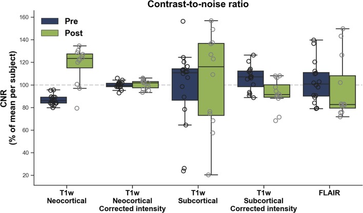
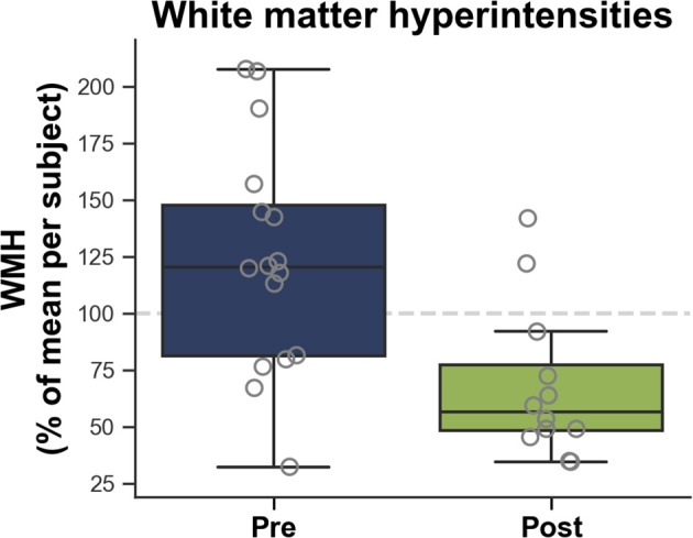
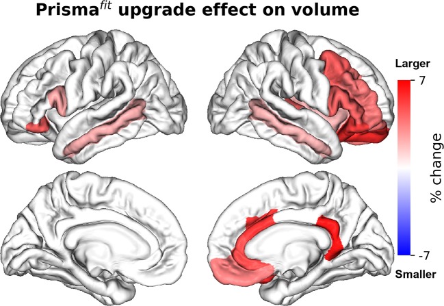
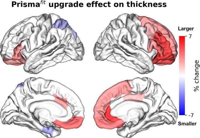
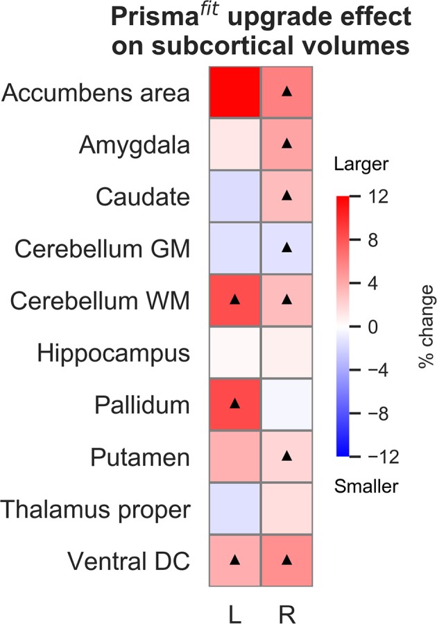
Major hardware/software changes to MRI platforms, either planned or unplanned, will almost invariably occur in longitudinal studies. Our objective was to assess the resulting variability on relevant imaging measurements in such context, specifically for three Siemens Healthcare Magnetom Trio upgrades to the Prisma platform. We report data acquired on three healthy volunteers scanned before and after three different platform upgrades. We assessed differences in image signal [contrast-to-noise ratio (CNR)] on T1-weighted images (T1w) and fluid-attenuated inversion recovery images (FLAIR); brain morphometry on T1w image; and small vessel disease (white matter hyperintensities; WMH) on FLAIR image. Prisma upgrade resulted in higher (30%) and more variable neocortical CNR and larger brain volume and thickness mainly in frontal areas. A significant relationship was observed between neocortical CNR and neocortical volume. For FLAIR images, no significant CNR difference was observed, but WMH volumes were significantly smaller (-68%) after Prisma upgrade, when compared to results on the Magnetom Trio. Together, these results indicate that Prisma upgrade significantly influenced image signal, brain morphometry measures and small vessel diseases measures and that these effects need to be taken into account when analyzing results from any longitudinal study undergoing similar changes.
在纵向研究中,磁共振成像(MRI)平台无论是计划内还是计划外的重大硬件/软件变更几乎总是会发生。我们的目的是评估在此种情况下相关成像测量结果产生的变异性,特别是针对西门子医疗公司的三款Magnetom Trio磁共振成像设备升级到Prisma平台的情况。我们报告了三名健康志愿者在三次不同平台升级前后扫描所获取的数据。我们评估了T1加权图像(T1w)和液体衰减反转恢复图像(FLAIR)上的图像信号差异[对比噪声比(CNR)];T1w图像上的脑形态测量;以及FLAIR图像上的小血管疾病(白质高信号;WMH)。Prisma平台升级后,新皮质的CNR更高(30%)且变异性更大,脑容量和厚度增加,主要集中在额叶区域。新皮质CNR与新皮质体积之间存在显著关系。对于FLAIR图像,未观察到显著的CNR差异,但与Magnetom Trio的结果相比,Prisma平台升级后WMH体积显著减小(-68%)。总之,这些结果表明Prisma平台升级显著影响了图像信号、脑形态测量指标和小血管疾病指标,在分析任何经历类似变更的纵向研究结果时都需要考虑这些影响。