Oregon Health & Science University, Layton Aging and Alzheimer's Disease Center, Neurology, USA; Oregon Health & Science University, Advanced Imaging Research Center, USA.
Oregon Health & Science University, Layton Aging and Alzheimer's Disease Center, Neurology, USA.
Neuroimage. 2019 Nov 15;202:116126. doi: 10.1016/j.neuroimage.2019.116126. Epub 2019 Aug 25.
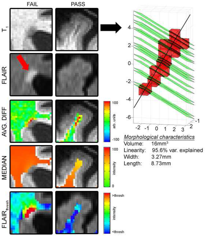
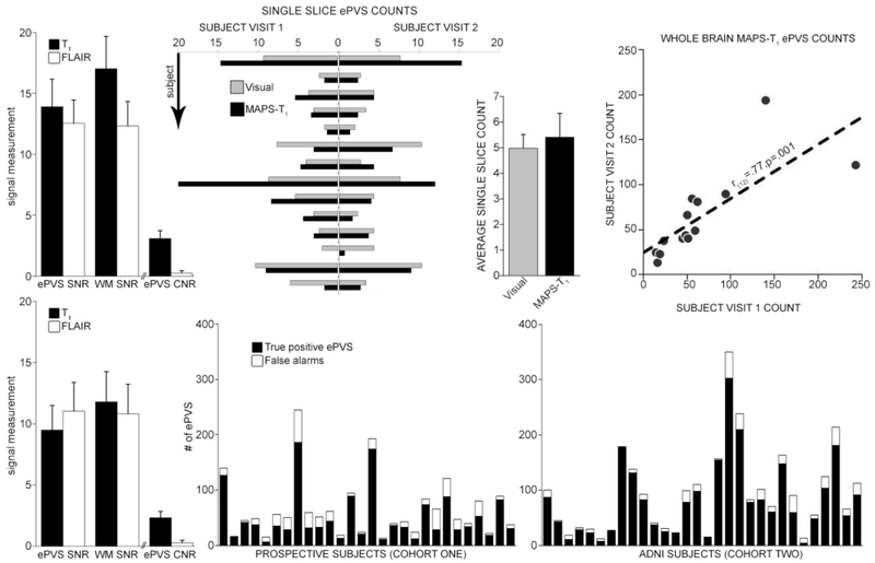
Recent interest in enlarged perivascular spaces (ePVS) in the brain, which can be visualized on MRI and appear isointense to cerebrospinal fluid on all sequence weightings, has resulted in the necessity of reliable algorithms for automated segmentation to allow for whole brain assessment of ePVS burden. However, several publicly available datasets do not contain sequences required for recently published algorithms. This prospective study presents a method for identification of enlarged perivascular spaces (ePVS) in white matter using 3T T and FLAIR MR imaging (MAPS-T), making the algorithm accessible to groups with valuable sets of limited data. The approach was applied identically to two datasets: 1) a repeated measurement in a dementia-free aged human population (N = 14), and 2) an aged sample of multisite ADNI datasets (N = 30). ePVS segmentation was accomplished by a stepwise local homogeneity search of white matter-masked T-weighted data, constrained by FLAIR hyperintensity, and further constrained by width, volume, and linearity measurements. Pearson's r was employed for statistical testing between visual (gold standard) assessment and repeated measures in cohort one. Visual ePVS counts were significantly correlated with MAPS-T (r = .72, P < .0001). Correlations between repeated measurements in cohort one were significant for both visual and automated methods in the single visually-rated slice (MAPS-T: r = .87, P < .0001, visual: (r = .86, P < .0001) and for whole brain assessment (MAPS-T: r = .77, P = .001). Results from each cohort were manually inspected and found to have positive predictive values of 77.5% and 87.5%, respectively. The approach described in this report is an important tool for detailed assessment of ePVS burden in white matter on routinely acquired MRI sequences.
最近人们对脑内扩大的血管周围间隙(ePVS)产生了兴趣,这些间隙在 MRI 上可以显示出来,在所有序列的加权图像上与脑脊液等信号强度,这使得需要可靠的自动分割算法来评估 ePVS 负荷。然而,一些现有的公开数据集不包含最近发表的算法所需的序列。本前瞻性研究提出了一种在 3T T 和 FLAIR MR 成像(MAPS-T)中识别脑白质内 ePVS 的方法,该方法使那些拥有有限数据集的研究组也可以使用这种算法。该方法被应用于两个数据集:1)无痴呆老年人群的重复测量(N=14),2)ADNI 多中心数据集的老年样本(N=30)。ePVS 分割通过对白质掩模 T 加权数据进行逐步局部同质性搜索来完成,该方法受 FLAIR 高信号的约束,进一步受宽度、体积和线性度测量的约束。在队列 1 中,采用 Pearson r 进行了视觉(金标准)评估与重复测量之间的统计学检验。视觉 ePVS 计数与 MAPS-T 显著相关(r=0.72,P<0.0001)。队列 1 中重复测量的相关性在视觉和自动方法中均显著,包括单张视觉评估切片(MAPS-T:r=0.87,P<0.0001,视觉:r=0.86,P<0.0001)和全脑评估(MAPS-T:r=0.77,P=0.001)。对每个队列的结果进行了手动检查,发现其阳性预测值分别为 77.5%和 87.5%。本报告中描述的方法是在常规采集的 MRI 序列上对白质内 ePVS 负荷进行详细评估的重要工具。