Division of Nephrology and Hypertension, Mayo Clinic, 200 First Street SW, Rochester, MN, 55905, USA.
Division of Maternal-Fetal Medicine, Department of Obstetrics and Gynecology, Mayo Clinic, Rochester, MN, USA.
Biol Sex Differ. 2019 Sep 14;10(1):49. doi: 10.1186/s13293-019-0263-5.
Preeclampsia is a pregnancy-specific hypertensive disorder characterized by impaired angiogenesis. We postulate that senescence of mesenchymal stem cells (MSC), multipotent cells with pro-angiogenic activities, is one of the mechanisms by which systemic inflammation exerts inhibitory effects on angiogenesis in preeclampsia.
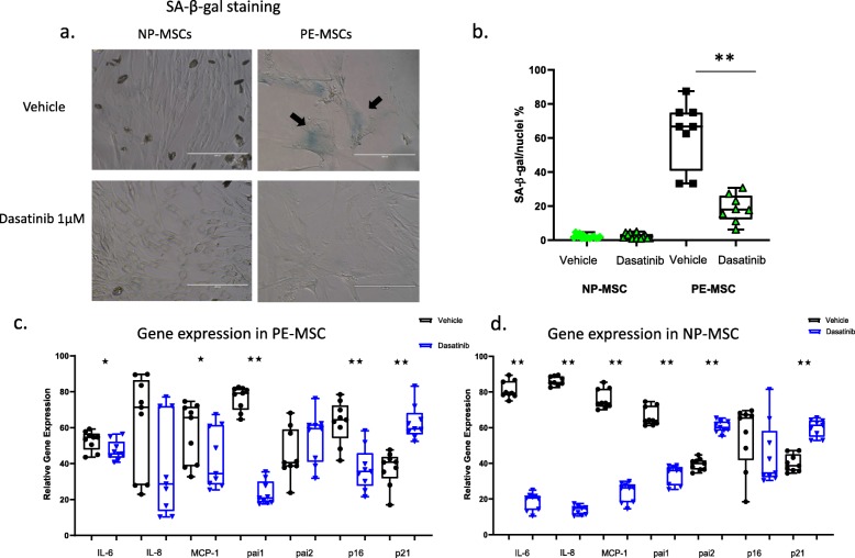
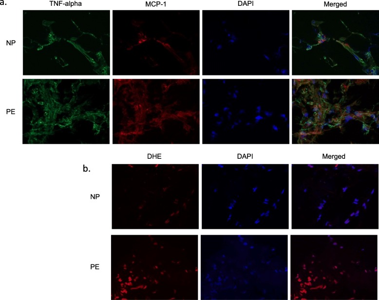
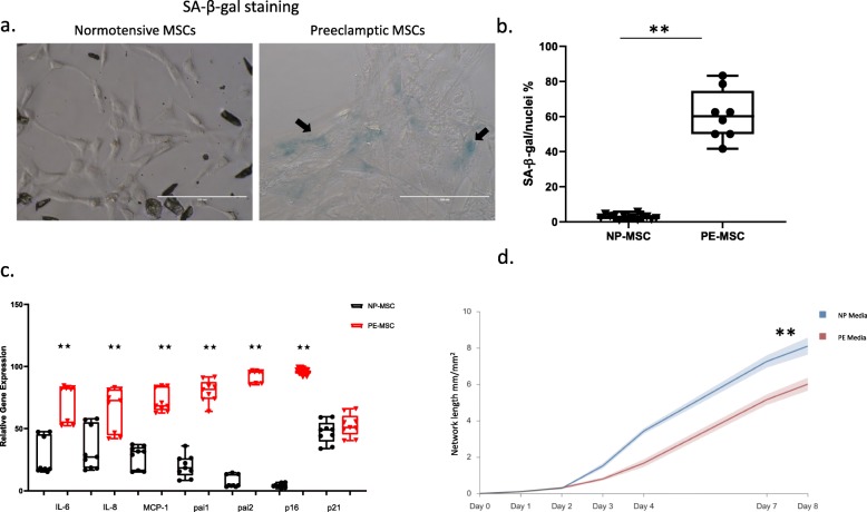
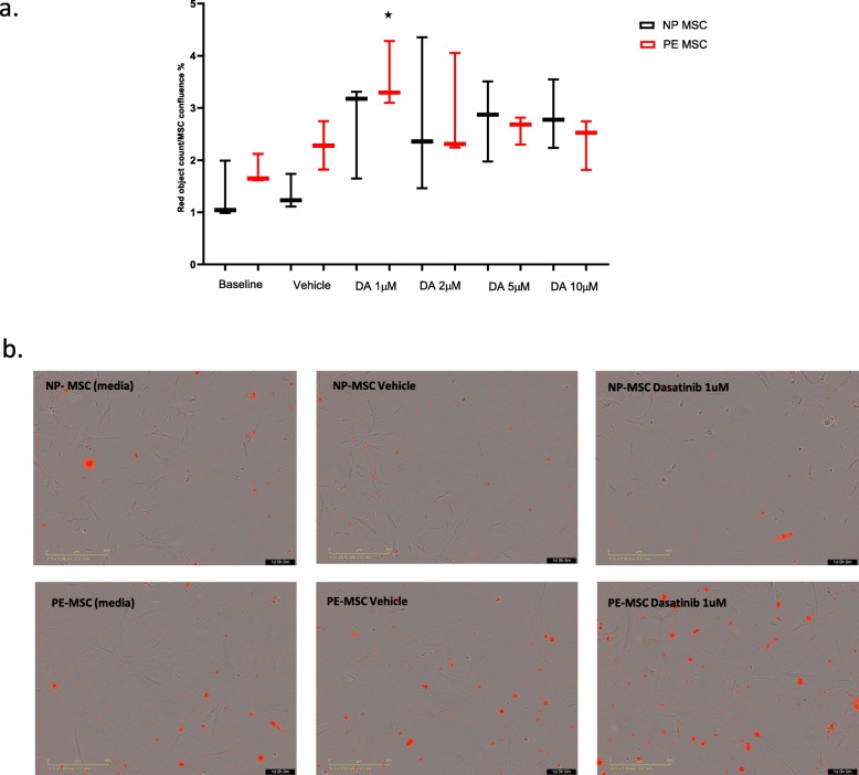
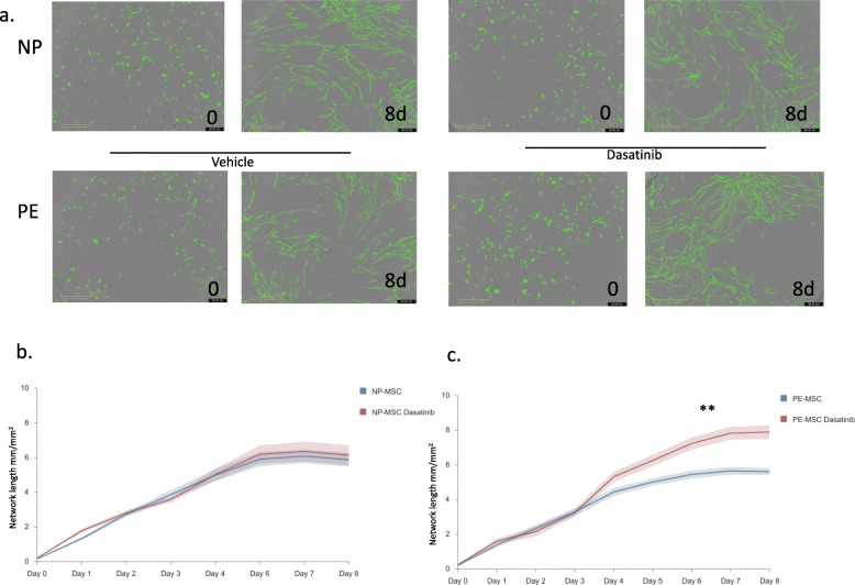
MSC were isolated from abdominal fat tissue explants removed during medically indicated C-sections from women with preeclampsia (PE-MSC, n = 10) and those with normotensive pregnancies (NP-MSC, n = 12). Sections of the frozen subcutaneous adipose tissue were assessed for inflammation by staining for tumor necrosis factor (TNF)-alpha and monocyte chemoattractant protein (MCP)-1. Viability, proliferation, and migration were compared between PE-MSC vs. NP-MSC. Apoptosis and angiogenesis were assayed before and after treatment with a senolytic agent (1 μM dasatinib) using the IncuCyte S3 Live-Cell Analysis System. Similarly, staining for senescence-associated beta galactosidase (SABG) and qPCR for gene expression of senescence markers, p16 and p21, as well as senescence-associated secretory phenotype (SASP) components, IL-6, IL-8, MCP-1, and PAI-1, were studied before and after treatment with dasatinib and compared between PE and NP.
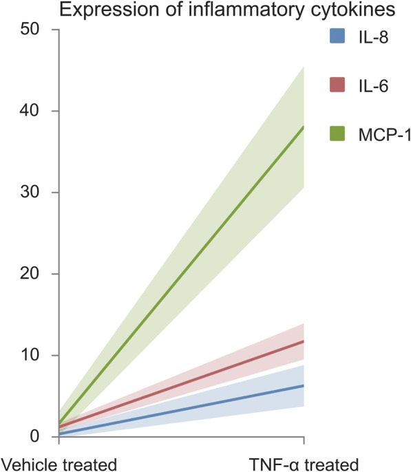
After in vitro exposure to TNF-alpha, MSC demonstrated upregulation of SASP components, including interleukins-6 and -8 and MCP-1. Staining of the subcutaneous adipose tissue sections revealed a greater inflammatory response in preeclampsia, based on the higher levels of both TNF-alpha and MCP-1 compared to normotensive pregnancies (p < 0.001 and 0.024, respectively). MSC isolated from PE demonstrated a lower percentage of live MSC cells (p = 0.012), lower proliferation (p = 0.005), and higher migration (p = 0.023). At baseline, PE-MSC demonstrated a senescent phenotype, reflected by more abundant staining for SABG (p < 0.001), upregulation of senescence markers and SASP components, as well as lower angiogenic potential (p < 0.001), compared to NP-MSC. Treatment with dasatinib increased significantly the number of apoptotic PE-MSC compared to NP-MSC (0.011 vs. 0.093) and decreased the gene expression of p16 and six SASP components. The mechanistic link between senescence and impaired angiogenesis in PE was confirmed by improved angiogenic potential of PE-MSC (p < 0.001) after dasatinib treatment.
Our data suggest that MSC senescence exerts inhibitory effects on angiogenesis in preeclampsia. Senolytic agents may offer the opportunity for mechanism-based therapies.
子痫前期是一种妊娠特异性高血压疾病,其特征为血管生成受损。我们假设,间充质干细胞(MSC)的衰老,这种具有促血管生成活性的多能细胞,是全身炎症对子痫前期血管生成产生抑制作用的机制之一。
从患有子痫前期(PE-MSC,n=10)和血压正常妊娠(NP-MSC,n=12)的女性剖宫产术中切除的腹部脂肪组织标本中分离 MSC。通过肿瘤坏死因子(TNF)-α和单核细胞趋化蛋白(MCP)-1染色评估冷冻皮下脂肪组织切片的炎症情况。比较 PE-MSC 与 NP-MSC 之间的活力、增殖和迁移情况。使用 IncuCyte S3 活细胞分析系统,在用 senolytic 剂(1μM 达沙替尼)处理前后,分别检测细胞凋亡和血管生成。同样,在使用达沙替尼处理前后,通过衰老相关β-半乳糖苷酶(SABG)染色和衰老标志物 p16 和 p21 以及衰老相关分泌表型(SASP)成分 IL-6、IL-8、MCP-1 和 PAI-1 的基因表达的 qPCR 研究,研究了 PE 和 NP 之间的差异。
MSC 在体外暴露于 TNF-α后,SASP 成分的表达上调,包括白细胞介素-6 和 -8 和 MCP-1。皮下脂肪组织切片染色显示,与血压正常妊娠相比,子痫前期的炎症反应更高,TNF-α和 MCP-1 水平均更高(p<0.001 和 0.024)。从 PE 分离的 MSC 表现出较低百分比的活 MSC 细胞(p=0.012),较低的增殖(p=0.005)和更高的迁移(p=0.023)。在基线时,PE-MSC 表现出衰老表型,这反映在 SABG 染色更为丰富(p<0.001),衰老标志物和 SASP 成分的上调,以及血管生成潜力降低(p<0.001),与 NP-MSC 相比。与 NP-MSC 相比,达沙替尼治疗后 PE-MSC 的凋亡细胞数量显著增加(0.011 对 0.093),并降低了 p16 和六个 SASP 成分的基因表达。PE-MSC 血管生成潜力改善证实了 PE 中衰老与血管生成受损之间的机制联系(p<0.001)。
我们的数据表明,MSC 衰老对子痫前期的血管生成产生抑制作用。衰老剂可能为基于机制的治疗提供机会。