• 文献检索
  • 文档翻译
  • 深度研究
  • 学术资讯
  • Suppr Zotero 插件Zotero 插件
  • 邀请有礼
  • 套餐&价格
  • 历史记录
应用&插件
Suppr Zotero 插件Zotero 插件浏览器插件Mac 客户端Windows 客户端微信小程序
定价
高级版会员购买积分包购买API积分包
服务
文献检索文档翻译深度研究API 文档MCP 服务
关于我们
关于 Suppr公司介绍联系我们用户协议隐私条款
关注我们

Suppr 超能文献

核心技术专利:CN118964589B侵权必究
粤ICP备2023148730 号-1Suppr @ 2026

文献检索

告别复杂PubMed语法,用中文像聊天一样搜索,搜遍4000万医学文献。AI智能推荐,让科研检索更轻松。

立即免费搜索

文件翻译

保留排版,准确专业,支持PDF/Word/PPT等文件格式,支持 12+语言互译。

免费翻译文档

深度研究

AI帮你快速写综述,25分钟生成高质量综述,智能提取关键信息,辅助科研写作。

立即免费体验

高强度超声脉冲序列致脑损伤的纵向功能评估

Longitudinal Functional Assessment of Brain Injury Induced by High-Intensity Ultrasound Pulse Sequences.

机构信息

Division of Biomedical Physics, Office of Science and Engineering Laboratories, Center for Devices and Radiological Health, Food and Drug Administration, Silver Spring, MD, USA.

Division of Neurological and Physical Medicine Devices, Office of Device Evaluation, Center for Devices and Radiological Health, Food and Drug Administration, Silver Spring, MD, USA.

出版信息

Sci Rep. 2019 Oct 29;9(1):15518. doi: 10.1038/s41598-019-51876-5.

DOI:10.1038/s41598-019-51876-5
PMID:31664091
原文链接:https://pmc.ncbi.nlm.nih.gov/articles/PMC6820547/
Abstract

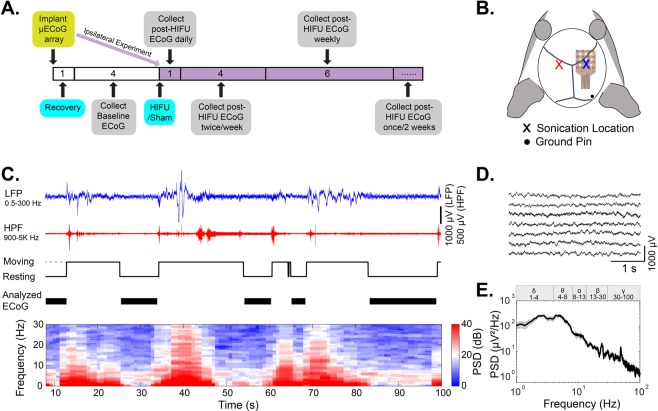
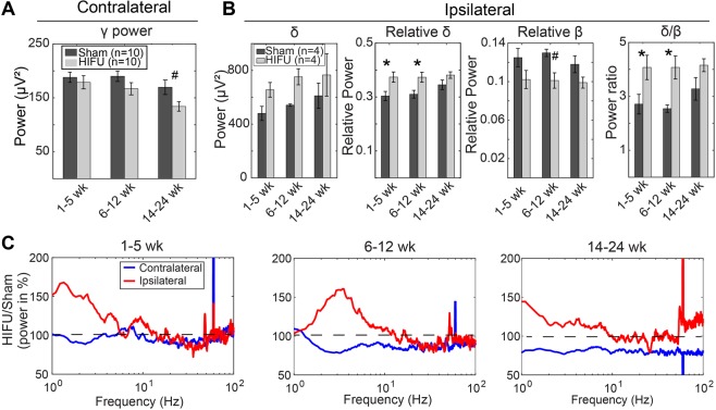
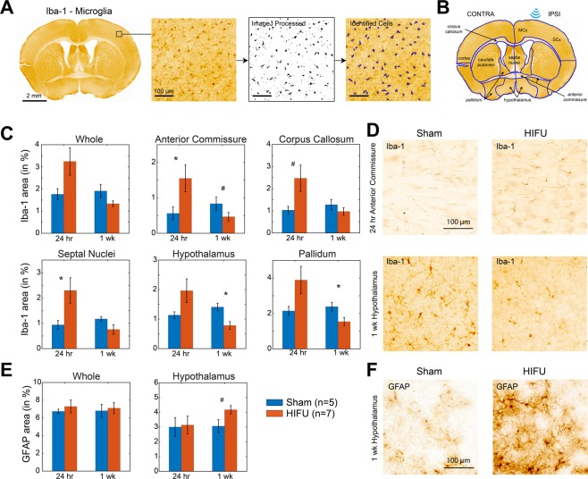
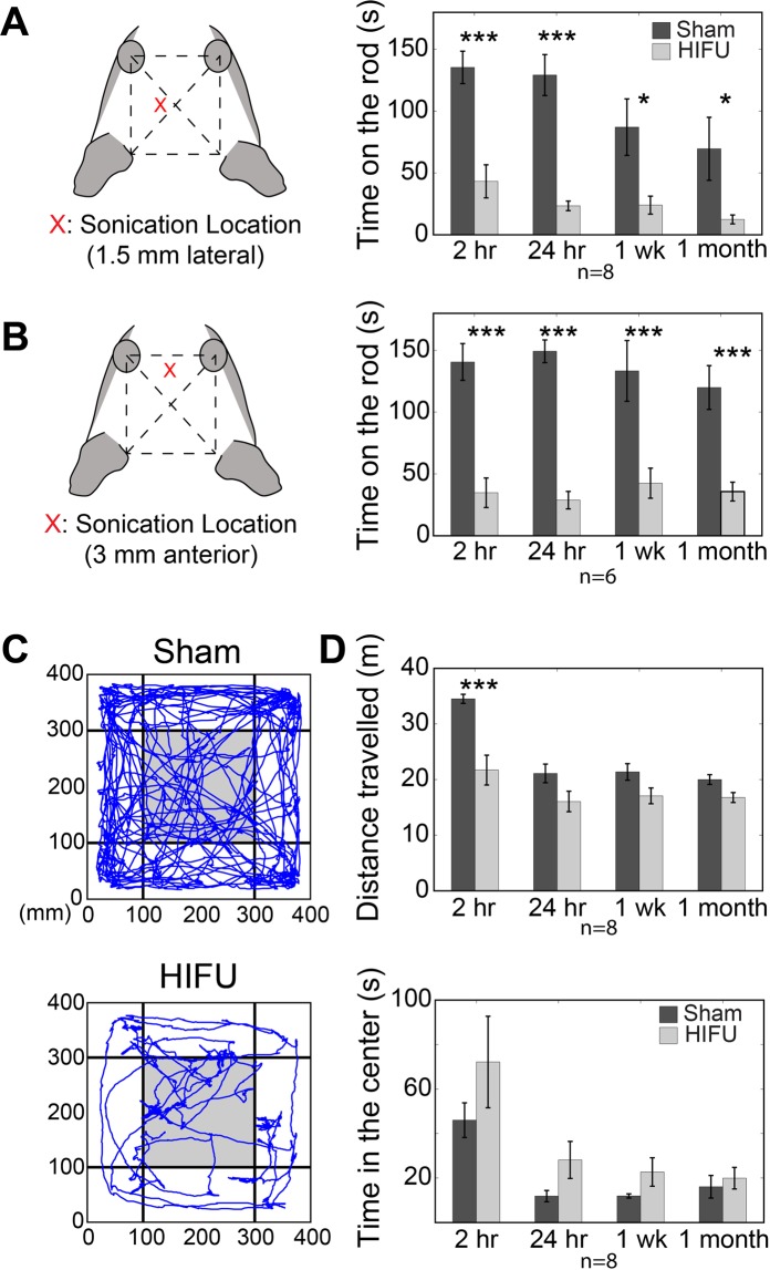
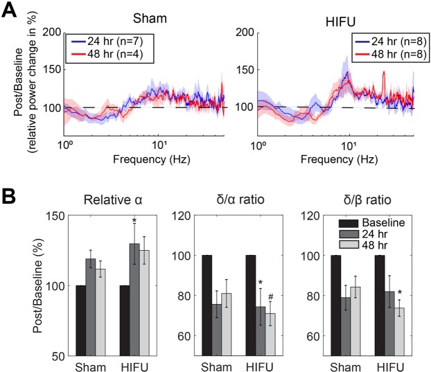
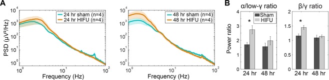
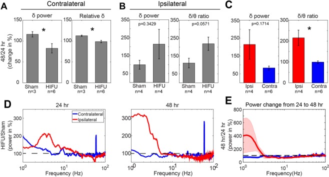
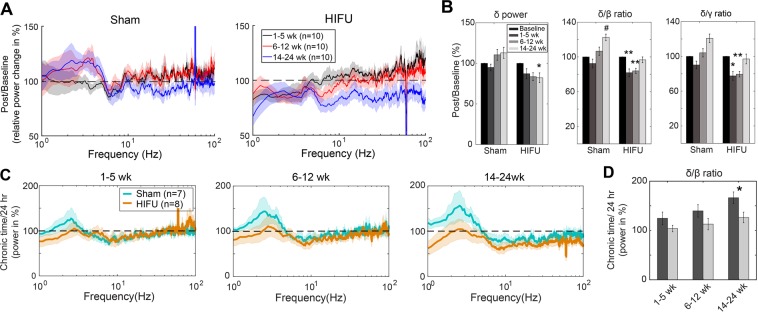
Exposure of the brain to high-intensity stress waves creates the potential for long-term functional deficits not related to thermal or cavitational damage. Possible sources of such exposure include overpressure from blast explosions or high-intensity focused ultrasound (HIFU). While current ultrasound clinical protocols do not normally produce long-term neurological deficits, the rapid expansion of potential therapeutic applications and ultrasound pulse-train protocols highlights the importance of establishing a safety envelope beyond which therapeutic ultrasound can cause neurological deficits not detectable by standard histological assessment for thermal and cavitational damage. In this study, we assessed the neuroinflammatory response, behavioral effects, and brain micro-electrocorticographic (µECoG) signals in mice following exposure to a train of transcranial pulses above normal clinical parameters. We found that the HIFU exposure induced a mild regional neuroinflammation not localized to the primary focal site, and impaired locomotor and exploratory behavior for up to 1 month post-exposure. In addition, low frequency (δ) and high frequency (β, γ) oscillations recorded by ECoG were altered at acute and chronic time points following HIFU application. ECoG signal changes on the hemisphere ipsilateral to HIFU exposure are of greater magnitude than the contralateral hemisphere, and persist for up to three months. These results are useful for describing the upper limit of transcranial ultrasound protocols, and the neurological sequelae of injury induced by high-intensity stress waves.

摘要

大脑暴露于高强度的应激波中,会产生与热或空化损伤无关的长期功能缺陷的可能性。这种暴露的可能来源包括爆炸冲击波的过压或高强度聚焦超声(HIFU)。虽然目前的超声临床方案通常不会产生长期的神经功能缺陷,但潜在治疗应用和超声脉冲序列方案的迅速扩展,突出了在治疗超声可能导致通过标准热和空化损伤组织学评估无法检测到的神经功能缺陷的安全范围之外建立安全界限的重要性。在这项研究中,我们评估了暴露于高于正常临床参数的一连串颅穿透脉冲后,小鼠的神经炎症反应、行为效应和脑微电皮质电图(µECoG)信号。我们发现,HIFU 暴露会引起轻度的区域性神经炎症,而不是局限于主要焦点部位,并在暴露后长达 1 个月的时间内损害运动和探索行为。此外,在 HIFU 应用后的急性和慢性时间点,ECoG 记录的低频(δ)和高频(β、γ)振荡发生了改变。与对侧半球相比,HIFU 暴露同侧半球的 ECoG 信号变化幅度更大,并且持续时间长达三个月。这些结果有助于描述颅穿透超声方案的上限,以及高强度应激波引起的损伤的神经后果。

https://cdn.ncbi.nlm.nih.gov/pmc/blobs/4d86/6820547/586942cdcc99/41598_2019_51876_Fig8_HTML.jpg
https://cdn.ncbi.nlm.nih.gov/pmc/blobs/4d86/6820547/c79960b1bd86/41598_2019_51876_Fig1_HTML.jpg
https://cdn.ncbi.nlm.nih.gov/pmc/blobs/4d86/6820547/a7638cebc4ae/41598_2019_51876_Fig2_HTML.jpg
https://cdn.ncbi.nlm.nih.gov/pmc/blobs/4d86/6820547/b4cdd6462514/41598_2019_51876_Fig3_HTML.jpg
https://cdn.ncbi.nlm.nih.gov/pmc/blobs/4d86/6820547/c119fcb68ab3/41598_2019_51876_Fig4_HTML.jpg
https://cdn.ncbi.nlm.nih.gov/pmc/blobs/4d86/6820547/ddc0bec50418/41598_2019_51876_Fig5_HTML.jpg
https://cdn.ncbi.nlm.nih.gov/pmc/blobs/4d86/6820547/7590c889aa86/41598_2019_51876_Fig6_HTML.jpg
https://cdn.ncbi.nlm.nih.gov/pmc/blobs/4d86/6820547/4620ede8e4ff/41598_2019_51876_Fig7_HTML.jpg
https://cdn.ncbi.nlm.nih.gov/pmc/blobs/4d86/6820547/586942cdcc99/41598_2019_51876_Fig8_HTML.jpg

相似文献

1
Longitudinal Functional Assessment of Brain Injury Induced by High-Intensity Ultrasound Pulse Sequences.高强度超声脉冲序列致脑损伤的纵向功能评估
Sci Rep. 2019 Oct 29;9(1):15518. doi: 10.1038/s41598-019-51876-5.
2
Application of high-intensity focused ultrasound to the study of mild traumatic brain injury.高强度聚焦超声在轻度创伤性脑损伤研究中的应用。
Ultrasound Med Biol. 2014 May;40(5):965-78. doi: 10.1016/j.ultrasmedbio.2013.11.023. Epub 2014 Jan 22.
3
Portable ultrasound-guided high-intensity focused ultrasound with functions for safe and rapid ablation: prospective clinical trial for uterine fibroids-short-term and long-term results.便携式超声引导高强度聚焦超声治疗,具有安全快速消融功能:子宫肌瘤的前瞻性临床试验——短期和长期结果。
Eur Radiol. 2020 Mar;30(3):1554-1563. doi: 10.1007/s00330-019-06468-2. Epub 2019 Nov 8.
4
Morphometric analysis of high-intensity focused ultrasound-induced lipolysis on cadaveric abdominal and thigh skin.高强度聚焦超声诱导尸体腹部和大腿皮肤脂肪分解的形态计量学分析
Lasers Med Sci. 2017 Jul;32(5):1143-1151. doi: 10.1007/s10103-017-2220-z. Epub 2017 Apr 27.
5
Investigation of HIFU-induced anti-tumor immunity in a murine tumor model.在小鼠肿瘤模型中对高强度聚焦超声(HIFU)诱导的抗肿瘤免疫的研究。
J Transl Med. 2007 Jul 11;5:34. doi: 10.1186/1479-5876-5-34.
6
Real-time monitoring of high-intensity focused ultrasound thermal therapy using the manifold learning method.使用流形学习方法对高强度聚焦超声热疗进行实时监测。
Ultrasound Med Biol. 2014 Dec;40(12):2841-50. doi: 10.1016/j.ultrasmedbio.2014.07.021.
7
Magnetic Resonance-Guided High-Intensity Focused Ultrasound (MR-HIFU): Technical Background and Overview of Current Clinical Applications (Part 1).磁共振引导高强度聚焦超声(MR-HIFU):技术背景与当前临床应用概述(第1部分)
Rofo. 2019 Jun;191(6):522-530. doi: 10.1055/a-0817-5645. Epub 2019 Jan 10.
8
Harmonic motion imaging for abdominal tumor detection and high-intensity focused ultrasound ablation monitoring: an in vivo feasibility study in a transgenic mouse model of pancreatic cancer.用于腹部肿瘤检测和高强度聚焦超声消融监测的谐波运动成像:在胰腺癌转基因小鼠模型中的体内可行性研究
IEEE Trans Ultrason Ferroelectr Freq Control. 2015 Sep;62(9):1662-73. doi: 10.1109/TUFFC.2015.007113.
9
High intensity focused ultrasound (HIFU) focal spot localization using harmonic motion imaging (HMI).使用谐波运动成像(HMI)进行高强度聚焦超声(HIFU)焦点定位。
Phys Med Biol. 2015 Aug 7;60(15):5911-24. doi: 10.1088/0031-9155/60/15/5911. Epub 2015 Jul 17.
10
Variations of bubble cavitation and temperature elevation during lesion formation by high-intensity focused ultrasound.高强度聚焦超声致伤过程中气泡空化和温升的变化。
J Acoust Soc Am. 2013 Aug;134(2):1683-94. doi: 10.1121/1.4812895.

引用本文的文献

1
Low frequency ultrasound elicits broad cortical responses inhibited by ketamine in mice.低频超声在小鼠中引发广泛的皮质反应,这些反应受到氯胺酮的抑制。
Commun Eng. 2024 Aug 27;3(1):120. doi: 10.1038/s44172-024-00269-2.
2
Amplitude- and frequency-dependent activation of layer II/III neurons by intracortical microstimulation.通过皮层内微刺激对II/III层神经元进行幅度和频率依赖性激活。
iScience. 2023 Oct 6;26(11):108140. doi: 10.1016/j.isci.2023.108140. eCollection 2023 Nov 17.
3
Evaluating Performance of EEG Data-Driven Machine Learning for Traumatic Brain Injury Classification.

本文引用的文献

1
Patients with Mild Traumatic Brain Injury Recruited from Both Hospital and Primary Care Settings: A Controlled Longitudinal Magnetic Resonance Imaging Study.从医院和初级保健机构招募轻度创伤性脑损伤患者:一项对照性纵向磁共振成像研究。
J Neurotrauma. 2019 Nov 15;36(22):3172-3182. doi: 10.1089/neu.2018.6360. Epub 2019 Jul 31.
2
Glial Fibrillary Acidic Protein (GFAP) Outperforms S100 Calcium-Binding Protein B (S100B) and Ubiquitin C-Terminal Hydrolase L1 (UCH-L1) as Predictor for Positive Computed Tomography of the Head in Trauma Subjects.胶质纤维酸性蛋白(GFAP)优于 S100 钙结合蛋白 B(S100B)和泛素 C 端水解酶 L1(UCH-L1),可作为创伤患者头部 CT 阳性的预测指标。
World Neurosurg. 2019 Aug;128:e434-e444. doi: 10.1016/j.wneu.2019.04.170. Epub 2019 May 1.
3
评估 EEG 数据驱动的机器学习在创伤性脑损伤分类中的性能。
IEEE Trans Biomed Eng. 2021 Nov;68(11):3205-3216. doi: 10.1109/TBME.2021.3062502. Epub 2021 Oct 19.
4
Longitudinal multimodal assessment of neurodegeneration and vascular remodeling correlated with signal degradation in chronic cortical silicon microelectrodes.慢性皮质硅微电极中神经退行性变和血管重塑的纵向多模态评估与信号降解的相关性
Neurophotonics. 2020 Jan;7(1):015004. doi: 10.1117/1.NPh.7.1.015004. Epub 2020 Jan 30.
Offline impact of transcranial focused ultrasound on cortical activation in primates.经颅聚焦超声对灵长类动物皮质激活的离线影响。
Elife. 2019 Feb 12;8:e40541. doi: 10.7554/eLife.40541.
4
Focused ultrasound thalamotomy location determines clinical benefits in patients with essential tremor.聚焦超声丘脑切开术的位置决定了原发性震颤患者的临床获益。
Brain. 2018 Dec 1;141(12):3405-3414. doi: 10.1093/brain/awy278.
5
Ultrasound Produces Extensive Brain Activation via a Cochlear Pathway.超声通过耳蜗通路产生广泛的脑激活。
Neuron. 2018 Aug 22;99(4):866. doi: 10.1016/j.neuron.2018.07.049.
6
Astrocyte activation and reactive gliosis-A new target in stroke?星形胶质细胞激活与反应性胶质增生——中风的新靶点?
Neurosci Lett. 2019 Jan 10;689:45-55. doi: 10.1016/j.neulet.2018.07.021. Epub 2018 Jul 17.
7
Ultrasonic Neuromodulation Causes Widespread Cortical Activation via an Indirect Auditory Mechanism.超声神经调节通过间接听觉机制引起广泛的皮层激活。
Neuron. 2018 Jun 6;98(5):1031-1041.e5. doi: 10.1016/j.neuron.2018.05.009. Epub 2018 May 24.
8
A Novel EEG Based Spectral Analysis of Persistent Brain Function Alteration in Athletes with Concussion History.基于 EEG 的运动员慢性脑震荡后持续性脑功能改变的频谱分析
Sci Rep. 2017 Dec 8;7(1):17221. doi: 10.1038/s41598-017-17414-x.
9
Epidermal Electrode Technology for Detecting Ultrasonic Perturbation of Sensory Brain Activity.表皮电极技术检测感觉性脑活动的超声干扰。
IEEE Trans Biomed Eng. 2018 Jun;65(6):1272-1280. doi: 10.1109/TBME.2017.2713647. Epub 2017 Aug 29.
10
Microglial Activation in Traumatic Brain Injury.创伤性脑损伤中的小胶质细胞激活
Front Aging Neurosci. 2017 Jun 28;9:208. doi: 10.3389/fnagi.2017.00208. eCollection 2017.