Department of Neurology, Massachusetts General Hospital, Charlestown, MA, 02129, USA.
Program in Neuroscience, Harvard Medical School, Boston, MA, 02115, USA.
Sci Rep. 2019 Nov 22;9(1):17387. doi: 10.1038/s41598-019-53554-y.
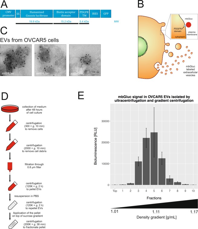
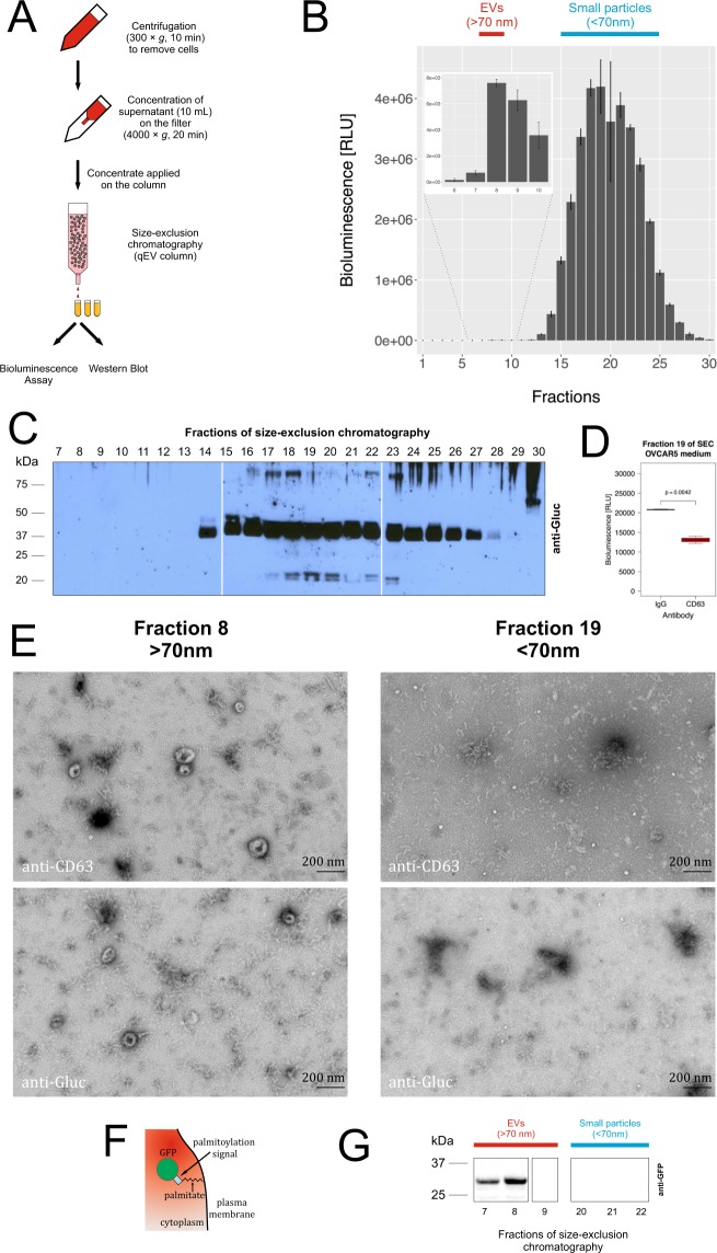
Extracellular vesicles (EVs) released by cells play a role in intercellular communication. Reporter and targeting proteins can be modified and exposed on the surface of EVs to investigate their half-life and biodistribution. A characterization of membrane-bound Gaussia luciferase (mbGluc) revealed that its signal was detected also in a form smaller than common EVs (<70 nm). We demonstrated that mbGluc initially exposed on the surface of EVs, likely undergoes proteolytic cleavage and processed fragments of the protein are released into the extracellular space in active form. Based on this observation, we developed a new assay to quantitatively track shedding of membrane proteins from the surface of EVs. We used this assay to show that ectodomain shedding in EVs is continuous and is mediated by specific proteases, e.g. metalloproteinases. Here, we present a novel tool to study membrane protein cleavage and release using both in vitro and in vivo models.
细胞释放的细胞外囊泡 (EVs) 在细胞间通讯中发挥作用。报告蛋白和靶向蛋白可以进行修饰并暴露在 EVs 表面,以研究其半衰期和生物分布。对膜结合型海肾荧光素酶 (mbGluc) 的特征分析表明,其信号也可以在比常见 EVs (<70nm) 更小的形式中检测到。我们证明 mbGluc 最初暴露在 EVs 表面,可能经历蛋白水解切割,并且该蛋白的加工片段以活性形式释放到细胞外空间。基于这一观察结果,我们开发了一种新的测定法来定量跟踪 EVs 表面膜蛋白的脱落。我们使用该测定法表明,EVs 中的外显肽脱落是连续的,并由特定的蛋白酶(例如金属蛋白酶)介导。在这里,我们提出了一种新的工具,可用于使用体外和体内模型研究膜蛋白的切割和释放。