• 文献检索
  • 文档翻译
  • 深度研究
  • 学术资讯
  • Suppr Zotero 插件Zotero 插件
  • 邀请有礼
  • 套餐&价格
  • 历史记录
应用&插件
Suppr Zotero 插件Zotero 插件浏览器插件Mac 客户端Windows 客户端微信小程序
定价
高级版会员购买积分包购买API积分包
服务
文献检索文档翻译深度研究API 文档MCP 服务
关于我们
关于 Suppr公司介绍联系我们用户协议隐私条款
关注我们

Suppr 超能文献

核心技术专利:CN118964589B侵权必究
粤ICP备2023148730 号-1Suppr @ 2026

文献检索

告别复杂PubMed语法,用中文像聊天一样搜索,搜遍4000万医学文献。AI智能推荐,让科研检索更轻松。

立即免费搜索

文件翻译

保留排版,准确专业,支持PDF/Word/PPT等文件格式,支持 12+语言互译。

免费翻译文档

深度研究

AI帮你快速写综述,25分钟生成高质量综述,智能提取关键信息,辅助科研写作。

立即免费体验

轴突周期性支架的超微结构显示出 actin 环的编织状组织。

Ultrastructure of the axonal periodic scaffold reveals a braid-like organization of actin rings.

机构信息

Sorbonne Université, INSERM, Institute of Myology, Centre of Research in Myology, UMRS 974, Paris, France.

Aix Marseille Université, CNRS, INP UMR7051, NeuroCyto, Marseille, France.

出版信息

Nat Commun. 2019 Dec 20;10(1):5803. doi: 10.1038/s41467-019-13835-6.

DOI:10.1038/s41467-019-13835-6
PMID:31862971
原文链接:https://pmc.ncbi.nlm.nih.gov/articles/PMC6925202/
Abstract

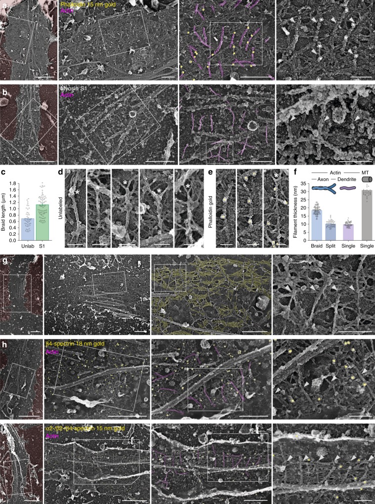
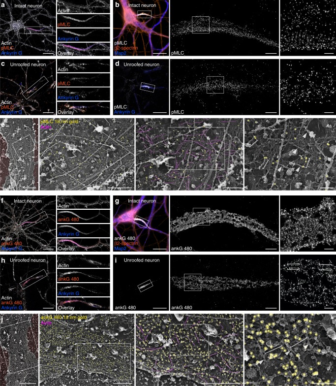
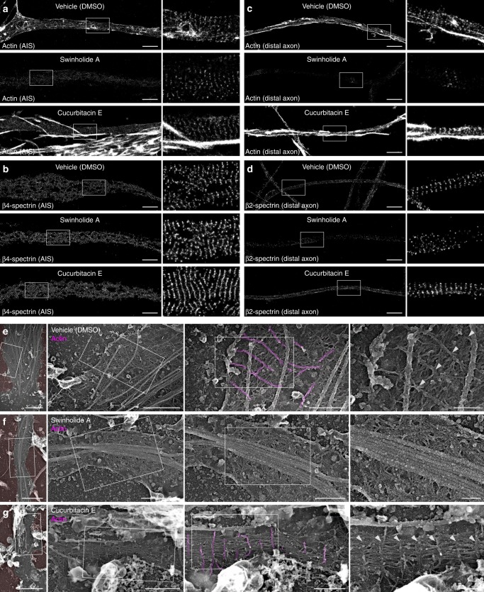
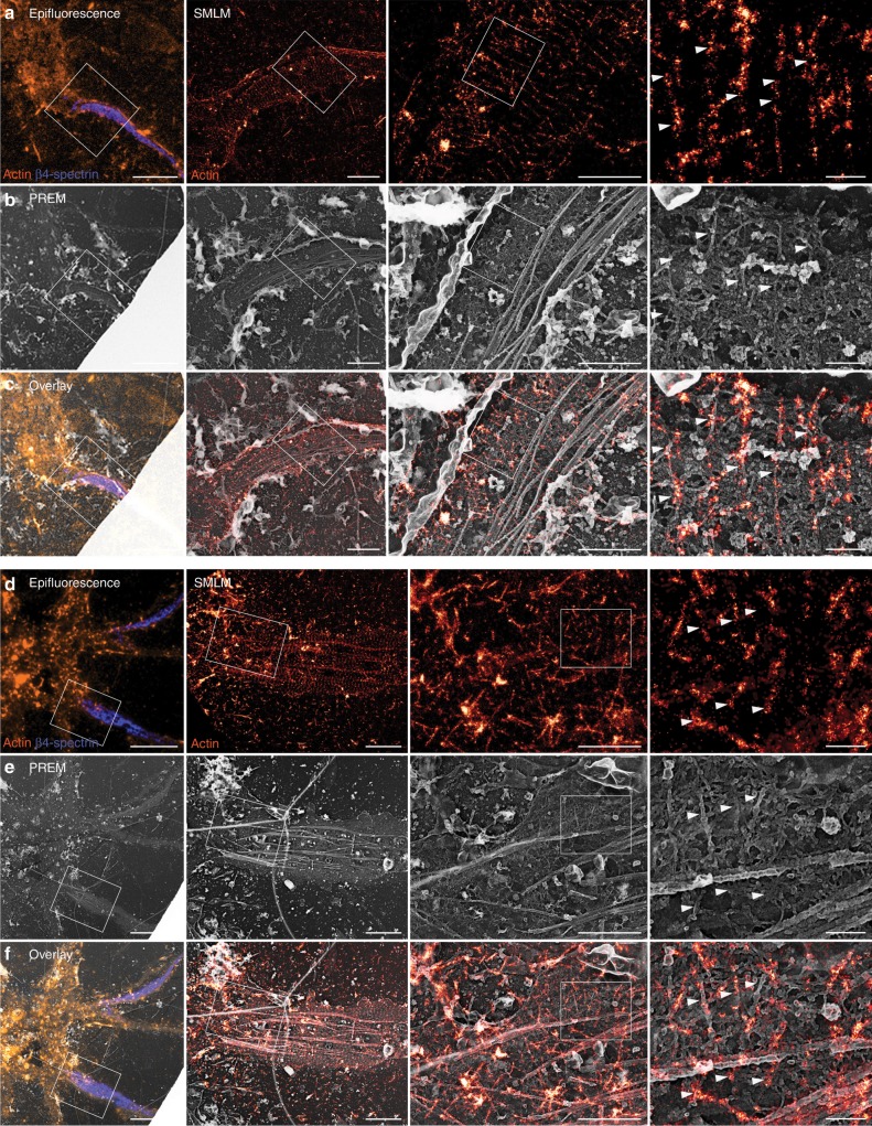
Recent super-resolution microscopy studies have unveiled a periodic scaffold of actin rings regularly spaced by spectrins under the plasma membrane of axons. However, ultrastructural details are unknown, limiting a molecular and mechanistic understanding of these enigmatic structures. Here, we combine platinum-replica electron and optical super-resolution microscopy to investigate the cortical cytoskeleton of axons at the ultrastructural level. Immunogold labeling and correlative super-resolution/electron microscopy allow us to unambiguously resolve actin rings as braids made of two long, intertwined actin filaments connected by a dense mesh of aligned spectrins. This molecular arrangement contrasts with the currently assumed model of actin rings made of short, capped actin filaments. Along the proximal axon, we resolved the presence of phospho-myosin light chain and the scaffold connection with microtubules via ankyrin G. We propose that braided rings explain the observed stability of the actin-spectrin scaffold and ultimately participate in preserving the axon integrity.

摘要

最近的超分辨率显微镜研究揭示了轴突质膜下肌动蛋白环周期性支架的存在,这些肌动蛋白环由 spectrins 规则间隔排列。然而,其超微结构细节尚不清楚,限制了对这些神秘结构的分子和机制理解。在这里,我们结合铂复制电子和光学超分辨率显微镜来研究轴突皮质细胞骨架的超微结构水平。免疫金标记和相关的超分辨率/电子显微镜使我们能够明确地将肌动蛋白环解析为由两条长的、相互交织的肌动蛋白丝连接而成的辫子,由排列整齐的 spectrins 组成密集的网格。这种分子排列与目前假设的由短的、加帽的肌动蛋白丝组成的肌动蛋白环模型形成对比。在近端轴突中,我们通过 ankyrin G 解析了磷酸化肌球蛋白轻链的存在和支架与微管的连接。我们提出,编织环解释了观察到的肌动蛋白 spectrin 支架的稳定性,并最终参与了轴突完整性的维持。

https://cdn.ncbi.nlm.nih.gov/pmc/blobs/b7b1/6925202/25608cf89945/41467_2019_13835_Fig6_HTML.jpg
https://cdn.ncbi.nlm.nih.gov/pmc/blobs/b7b1/6925202/5e42e3b848fa/41467_2019_13835_Fig1_HTML.jpg
https://cdn.ncbi.nlm.nih.gov/pmc/blobs/b7b1/6925202/46dc911e939a/41467_2019_13835_Fig2_HTML.jpg
https://cdn.ncbi.nlm.nih.gov/pmc/blobs/b7b1/6925202/a7bb6a05a57c/41467_2019_13835_Fig3_HTML.jpg
https://cdn.ncbi.nlm.nih.gov/pmc/blobs/b7b1/6925202/d18ae82a10cf/41467_2019_13835_Fig4_HTML.jpg
https://cdn.ncbi.nlm.nih.gov/pmc/blobs/b7b1/6925202/64958e8079a5/41467_2019_13835_Fig5_HTML.jpg
https://cdn.ncbi.nlm.nih.gov/pmc/blobs/b7b1/6925202/25608cf89945/41467_2019_13835_Fig6_HTML.jpg

相似文献

1
Ultrastructure of the axonal periodic scaffold reveals a braid-like organization of actin rings.轴突周期性支架的超微结构显示出 actin 环的编织状组织。
Nat Commun. 2019 Dec 20;10(1):5803. doi: 10.1038/s41467-019-13835-6.
2
Actin, spectrin, and associated proteins form a periodic cytoskeletal structure in axons.肌动蛋白、血影蛋白和相关蛋白在轴突中形成周期性细胞骨架结构。
Science. 2013 Jan 25;339(6118):452-6. doi: 10.1126/science.1232251. Epub 2012 Dec 13.
3
Modeling of the axon membrane skeleton structure and implications for its mechanical properties.轴突膜骨架结构建模及其力学性能研究
PLoS Comput Biol. 2017 Feb 27;13(2):e1005407. doi: 10.1371/journal.pcbi.1005407. eCollection 2017 Feb.
4
Cytoskeleton: axons earn their stripes.细胞骨架:轴突获得条纹。
Curr Biol. 2013 Mar 4;23(5):R197-8. doi: 10.1016/j.cub.2013.01.050.
5
Putting the axonal periodic scaffold in order.整理轴突周期性支架。
Curr Opin Neurobiol. 2021 Aug;69:33-40. doi: 10.1016/j.conb.2020.12.015. Epub 2021 Jan 12.
6
Structural Basis Underlying Strong Interactions between Ankyrins and Spectrins.锚蛋白与血影蛋白相互作用的结构基础。
J Mol Biol. 2020 Jun 12;432(13):3838-3850. doi: 10.1016/j.jmb.2020.04.023. Epub 2020 Apr 27.
7
Structural organization of the actin-spectrin-based membrane skeleton in dendrites and soma of neurons.神经元树突和胞体中肌动蛋白-血影蛋白为基础的膜骨架的结构组织。
Proc Natl Acad Sci U S A. 2017 Aug 8;114(32):E6678-E6685. doi: 10.1073/pnas.1705043114. Epub 2017 Jul 24.
8
Remodeling of the Actin/Spectrin Membrane-associated Periodic Skeleton, Growth Cone Collapse and F-Actin Decrease during Axonal Degeneration.轴突变性过程中肌动蛋白/血影蛋白膜相关周期骨架的重构、生长锥塌陷和 F-肌动蛋白减少。
Sci Rep. 2018 Feb 14;8(1):3007. doi: 10.1038/s41598-018-21232-0.
9
Nanoscale Architecture of the Axon Initial Segment Reveals an Organized and Robust Scaffold.轴突起始段的纳米级结构揭示了一种有序而坚固的支架。
Cell Rep. 2015 Dec 29;13(12):2781-93. doi: 10.1016/j.celrep.2015.11.051. Epub 2015 Dec 17.
10
Axon initial segment cytoskeleton comprises a multiprotein submembranous coat containing sparse actin filaments.轴突起始段细胞骨架由包含稀疏肌动蛋白丝的多蛋白亚膜包被组成。
J Cell Biol. 2014 Apr 14;205(1):67-81. doi: 10.1083/jcb.201401045. Epub 2014 Apr 7.

引用本文的文献

1
A correlation-based tool for quantifying membrane periodic skeleton associated periodicity.一种基于相关性的工具,用于量化膜相关周期性骨架的周期性。
Front Neuroinform. 2025 Aug 22;19:1628538. doi: 10.3389/fninf.2025.1628538. eCollection 2025.
2
The membrane skeleton is constitutively remodeled in neurons by calcium signaling.在神经元中,膜骨架通过钙信号传导持续重塑。
Science. 2025 Aug 7;389(6760):eadn6712. doi: 10.1126/science.adn6712.
3
Potassium channel clustering: mechanisms shaping axonal excitability.钾通道聚集:塑造轴突兴奋性的机制

本文引用的文献

1
Tropomyosin Tpm3.1 Is Required to Maintain the Structure and Function of the Axon Initial Segment.原肌球蛋白Tpm3.1是维持轴突起始段结构和功能所必需的。
iScience. 2020 May 22;23(5):101053. doi: 10.1016/j.isci.2020.101053. Epub 2020 Apr 12.
2
Axonal transport and neurological disease.轴突运输与神经疾病。
Nat Rev Neurol. 2019 Dec;15(12):691-703. doi: 10.1038/s41582-019-0257-2. Epub 2019 Sep 26.
3
Membrane-associated periodic skeleton is a signaling platform for RTK transactivation in neurons.膜相关周期骨架是神经元中 RTK 转激活的信号平台。
Front Cell Neurosci. 2025 Jul 1;19:1627517. doi: 10.3389/fncel.2025.1627517. eCollection 2025.
4
Nonmuscle myosin II regulates presynaptic actin and neuronal mechanobiology in Drosophila.非肌肉肌球蛋白II调节果蝇突触前肌动蛋白和神经元力学生物学。
J Cell Biol. 2025 Sep 1;224(9). doi: 10.1083/jcb.202501211. Epub 2025 Jul 11.
5
Super-resolution imaging of the neuronal cytoskeleton.神经元细胞骨架的超分辨率成像。
Npj Imaging. 2024 Dec 4;2(1):50. doi: 10.1038/s44303-024-00054-y.
6
Structural Repetition Detector for multi-scale quantitative mapping of molecular complexes through microscopy.用于通过显微镜对分子复合物进行多尺度定量映射的结构重复检测器。
Nat Commun. 2025 Jul 1;16(1):5767. doi: 10.1038/s41467-025-60709-1.
7
Myosin 2 - A general contractor for the cytoskeleton.肌球蛋白2——细胞骨架的总承包商。
Curr Opin Cell Biol. 2025 Jun;94:102522. doi: 10.1016/j.ceb.2025.102522. Epub 2025 May 3.
8
Paralemmin-1 controls the nanoarchitecture of the neuronal submembrane cytoskeleton.Paralemmin-1蛋白控制着神经元亚膜细胞骨架的纳米结构。
Sci Adv. 2025 Mar 7;11(10):eadt3724. doi: 10.1126/sciadv.adt3724.
9
Membrane mechanics dictate axonal pearls-on-a-string morphology and function.膜力学决定轴突上珍珠串样形态和功能。
Nat Neurosci. 2025 Jan;28(1):49-61. doi: 10.1038/s41593-024-01813-1. Epub 2024 Dec 2.
10
Unveiling the cell biology of hippocampal neurons with dendritic axon origin.揭示具有树突轴突起源的海马神经元的细胞生物学。
J Cell Biol. 2025 Jan 6;224(1). doi: 10.1083/jcb.202403141. Epub 2024 Nov 4.
Science. 2019 Aug 30;365(6456):929-934. doi: 10.1126/science.aaw5937.
4
About samples, giving examples: Optimized Single Molecule Localization Microscopy.关于样本,举例来说:优化单分子定位显微镜。
Methods. 2020 Mar 1;174:100-114. doi: 10.1016/j.ymeth.2019.05.008. Epub 2019 May 10.
5
Sub-membranous actin rings in the axon initial segment are resistant to the action of latrunculin.轴突起始段的亚膜肌动蛋白环能抵抗 latrunculin 的作用。
Biol Chem. 2019 Aug 27;400(9):1141-1146. doi: 10.1515/hsz-2019-0111.
6
Visualizing and discovering cellular structures with super-resolution microscopy.用超分辨率显微镜可视化和发现细胞结构。
Science. 2018 Aug 31;361(6405):880-887. doi: 10.1126/science.aau1044. Epub 2018 Aug 30.
7
Exploring cytoskeletal diversity in neurons.探索神经元中的细胞骨架多样性。
Science. 2018 Jul 20;361(6399):231-232. doi: 10.1126/science.aat5992.
8
Actin Assemblies in the Axon Shaft - some Open Questions.轴突中的肌动蛋白组装 - 一些未解决的问题。
Curr Opin Neurobiol. 2018 Aug;51:163-167. doi: 10.1016/j.conb.2018.06.012. Epub 2018 Jul 10.
9
The Actin/Spectrin Membrane-Associated Periodic Skeleton in Neurons.神经元中的肌动蛋白/血影蛋白膜相关周期性骨架
Front Synaptic Neurosci. 2018 May 23;10:10. doi: 10.3389/fnsyn.2018.00010. eCollection 2018.
10
Pleomorphic linkers as ubiquitous structural organizers of vesicles in axons.多形连接子作为轴突中囊泡的普遍结构组织者。
PLoS One. 2018 Jun 4;13(6):e0197886. doi: 10.1371/journal.pone.0197886. eCollection 2018.