• 文献检索
  • 文档翻译
  • 深度研究
  • 学术资讯
  • Suppr Zotero 插件Zotero 插件
  • 邀请有礼
  • 套餐&价格
  • 历史记录
应用&插件
Suppr Zotero 插件Zotero 插件浏览器插件Mac 客户端Windows 客户端微信小程序
定价
高级版会员购买积分包购买API积分包
服务
文献检索文档翻译深度研究API 文档MCP 服务
关于我们
关于 Suppr公司介绍联系我们用户协议隐私条款
关注我们

Suppr 超能文献

核心技术专利:CN118964589B侵权必究
粤ICP备2023148730 号-1Suppr @ 2026

文献检索

告别复杂PubMed语法,用中文像聊天一样搜索,搜遍4000万医学文献。AI智能推荐,让科研检索更轻松。

立即免费搜索

文件翻译

保留排版,准确专业,支持PDF/Word/PPT等文件格式,支持 12+语言互译。

免费翻译文档

深度研究

AI帮你快速写综述,25分钟生成高质量综述,智能提取关键信息,辅助科研写作。

立即免费体验

蓝光诱导培养神经元中的基因表达改变是与神经元培养基的光毒性相互作用的结果。

Blue Light-Induced Gene Expression Alterations in Cultured Neurons Are the Result of Phototoxic Interactions with Neuronal Culture Media.

机构信息

Department of Neurobiology and Evelyn F. McKnight Brain Institute, University of Alabama at Birmingham, Birmingham, AL 35294.

Behavioral Neuroscience Research Branch, Intramural Research Program, National Institute on Drug Abuse, NIH/DHHS, Baltimore, MD 21224.

出版信息

eNeuro. 2020 Jan 6;7(1). doi: 10.1523/ENEURO.0386-19.2019. Print 2020 Jan/Feb.

DOI:10.1523/ENEURO.0386-19.2019
PMID:31879366
原文链接:https://pmc.ncbi.nlm.nih.gov/articles/PMC6946540/
Abstract

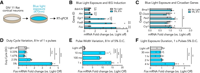
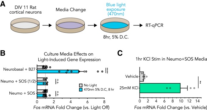
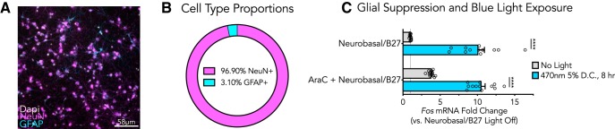
Blue wavelength light is used as an optical actuator in numerous optogenetic technologies employed in neuronal systems. However, the potential side effects of blue light in neurons has not been thoroughly explored, and recent reports suggest that neuronal exposure to blue light can induce transcriptional alterations and Here, we examined the effects of blue wavelength light in cultured primary rat cortical cells. Exposure to blue light (470 nm) resulted in upregulation of several immediate early genes (IEGs) traditionally used as markers of neuronal activity, including and , but did not alter the expression of circadian clock genes , , , , or IEG expression was increased following 4 h of 5% duty cycle light exposure, and IEG induction was not dependent on light pulse width. Elevated levels of blue light exposure induced a loss of cell viability , suggestive of overt phototoxicity. Induction of IEGs by blue light was maintained in cortical cultures treated with AraC to block glial proliferation, indicating that induction occurred selectively in postmitotic neurons. Importantly, changes in gene expression induced by blue wavelength light were prevented when cultures were maintained in a photoinert media supplemented with a photostable neuronal supplement instead of commonly utilized neuronal culture media and supplements. Together, these findings suggest that light-induced gene expression alterations observed stem from a phototoxic interaction between commonly used media and neurons, and offer a solution to prevent this toxicity when using photoactivatable technology .

摘要

蓝色波长光被用作许多用于神经元系统的光遗传学技术中的光学致动器。然而,神经元中蓝光的潜在副作用尚未得到彻底探索,最近的报告表明,神经元暴露于蓝光会诱导转录改变。在这里,我们检查了培养的原代大鼠皮质细胞中蓝色波长光的作用。暴露于蓝光(470nm)导致几个即时早期基因(IEGs)的上调,这些基因通常被用作神经元活动的标志物,包括 和 ,但不改变生物钟基因的表达 、 、 、 或 。IEG 的表达在 5%占空比光暴露 4 小时后增加,IEG 诱导不依赖于光脉冲宽度。高水平的蓝光暴露诱导细胞活力丧失,提示明显的光毒性。蓝光诱导的 IEG 表达在用 AraC 处理以阻断神经胶质增殖的皮质培养物中得以维持,表明诱导选择性地发生在有丝分裂后的神经元中。重要的是,当培养物在补充有光稳定神经元补充剂而不是常用神经元培养基和补充剂的光惰性培养基中维持时,蓝色波长光诱导的基因表达变化被阻止。这些发现表明,观察到的光诱导基因表达改变源于常用培养基和神经元之间的光毒性相互作用,并提供了一种在使用光激活技术时防止这种毒性的解决方案。

https://cdn.ncbi.nlm.nih.gov/pmc/blobs/f540/6946540/e5ff1699d8bb/enu9991931630004.jpg
https://cdn.ncbi.nlm.nih.gov/pmc/blobs/f540/6946540/4bc772fbd8bc/enu9991931630001.jpg
https://cdn.ncbi.nlm.nih.gov/pmc/blobs/f540/6946540/ac6fba07f04a/enu9991931630002.jpg
https://cdn.ncbi.nlm.nih.gov/pmc/blobs/f540/6946540/0020814b6f7b/enu9991931630003.jpg
https://cdn.ncbi.nlm.nih.gov/pmc/blobs/f540/6946540/e5ff1699d8bb/enu9991931630004.jpg

相似文献

1
Blue Light-Induced Gene Expression Alterations in Cultured Neurons Are the Result of Phototoxic Interactions with Neuronal Culture Media.蓝光诱导培养神经元中的基因表达改变是与神经元培养基的光毒性相互作用的结果。
eNeuro. 2020 Jan 6;7(1). doi: 10.1523/ENEURO.0386-19.2019. Print 2020 Jan/Feb.
2
Blue Light Increases Neuronal Activity-Regulated Gene Expression in the Absence of Optogenetic Proteins.蓝光在没有光遗传学蛋白的情况下增加神经元活动调节的基因表达。
eNeuro. 2019 Sep 18;6(5). doi: 10.1523/ENEURO.0085-19.2019. Print 2019 Sep/Oct.
3
Identification of optogenetically activated striatal medium spiny neurons by Npas4 expression.通过 Npas4 表达鉴定光遗传学激活的纹状体中型棘突神经元。
PLoS One. 2012;7(12):e52783. doi: 10.1371/journal.pone.0052783. Epub 2012 Dec 26.
4
Photoperiodic modulation of clock gene expression in the avian premammillary nucleus.光周期对禽类前乳头核时钟基因表达的调制。
J Neuroendocrinol. 2010 Feb;22(2):119-28. doi: 10.1111/j.1365-2826.2009.01942.x. Epub 2009 Dec 4.
5
BrainPhys neuronal medium optimized for imaging and optogenetics in vitro.BrainPhys 神经元培养基,优化用于体外成像和光遗传学。
Nat Commun. 2020 Nov 3;11(1):5550. doi: 10.1038/s41467-020-19275-x.
6
The role of Jun transcription factor expression and phosphorylation in neuronal differentiation, neuronal cell death, and plastic adaptations in vivo.Jun转录因子表达和磷酸化在体内神经元分化、神经元细胞死亡及可塑性适应中的作用。
Cell Mol Neurobiol. 1994 Oct;14(5):487-505. doi: 10.1007/BF02088833.
7
Daily rhythm of spontaneous immediate-early gene expression in the rat suprachiasmatic nucleus.大鼠视交叉上核中自发即刻早期基因表达的日节律。
J Biol Rhythms. 1999 Aug;14(4):275-80. doi: 10.1177/074873099129000687.
8
Ischemia-induced CA1 neuronal death is preceded by elevated FosB and Jun expression and reduced NGFI-A and JunB levels.在缺血诱导的CA1神经元死亡之前,FosB和Jun表达升高,而NGFI-A和JunB水平降低。
Brain Res Mol Brain Res. 1998 May;56(1-2):146-61. doi: 10.1016/s0169-328x(98)00039-4.
9
Blue light exposure in vitro causes toxicity to trigeminal neurons and glia through increased superoxide and hydrogen peroxide generation.体外蓝光照射会通过增加超氧化物和过氧化氢的产生,导致三叉神经神经元和神经胶质细胞毒性。
Free Radic Biol Med. 2019 Feb 1;131:27-39. doi: 10.1016/j.freeradbiomed.2018.11.029. Epub 2018 Nov 27.
10
Temporal expression of clock genes in central and peripheral tissues of spotted munia under varying light conditions: Evidence for circadian regulation of daily physiology in a non-photoperiodic circannual songbird species.斑文鸟中枢和外周组织中生物钟基因在不同光照条件下的时序表达:非光周期的年周期鸣禽物种日常生理节律调控的证据
Chronobiol Int. 2018 May;35(5):617-632. doi: 10.1080/07420528.2017.1422742. Epub 2018 Jan 25.

引用本文的文献

1
Blue Light-Induced, Dosed Protein Expression of Active BDNF in Human Cells Using the Optogenetic CRY2/CIB System.利用光遗传学CRY2/CIB系统在人细胞中蓝光诱导的活性脑源性神经营养因子的定量蛋白表达
Biotechnol J. 2024 Dec;19(12):e202400384. doi: 10.1002/biot.202400384.
2
Distinct roles of Bdnf I and Bdnf IV transcript variant expression in hippocampal neurons.脑源性神经营养因子I和脑源性神经营养因子IV转录变体表达在海马神经元中的不同作用。
Hippocampus. 2024 May;34(5):218-229. doi: 10.1002/hipo.23600. Epub 2024 Feb 16.
3
Single cell optogenetic stimulation by two-photon excitation fluorescence transfer.

本文引用的文献

1
Blue Light Increases Neuronal Activity-Regulated Gene Expression in the Absence of Optogenetic Proteins.蓝光在没有光遗传学蛋白的情况下增加神经元活动调节的基因表达。
eNeuro. 2019 Sep 18;6(5). doi: 10.1523/ENEURO.0085-19.2019. Print 2019 Sep/Oct.
2
Pharmacologic inhibition of LIMK1 provides dendritic spine resilience against β-amyloid.抑制 LIMK1 的药理作用可增强树突棘对β-淀粉样蛋白的抵抗力。
Sci Signal. 2019 Jun 25;12(587):eaaw9318. doi: 10.1126/scisignal.aaw9318.
3
Thermal constraints on in vivo optogenetic manipulations.体内光遗传学操作的热限制。
通过双光子激发荧光转移进行单细胞光遗传学刺激。
iScience. 2023 Sep 9;26(10):107857. doi: 10.1016/j.isci.2023.107857. eCollection 2023 Oct 20.
4
Inducible CRISPR Epigenome Systems Mimic Cocaine Induced Bidirectional Regulation of Nab2 and Egr3.诱导型 CRISPR 表观基因组系统模拟可卡因诱导的 Nab2 和 Egr3 的双向调节。
J Neurosci. 2023 Mar 29;43(13):2242-2259. doi: 10.1523/JNEUROSCI.1802-22.2022. Epub 2023 Feb 27.
5
Chimeric GPCRs mimic distinct signaling pathways and modulate microglia responses.嵌合 G 蛋白偶联受体模拟不同的信号通路,并调节小胶质细胞的反应。
Nat Commun. 2022 Aug 15;13(1):4728. doi: 10.1038/s41467-022-32390-1.
6
An Improved CRISPR/dCas9 Interference Tool for Neuronal Gene Suppression.一种用于神经元基因抑制的改进型CRISPR/dCas9干扰工具。
Front Genome Ed. 2020 Sep 15;2:9. doi: 10.3389/fgeed.2020.00009. eCollection 2020.
7
Dose-dependent threshold illumination for non-invasive time-lapse fluorescence imaging of live cells.活细胞非侵入性延时荧光成像的剂量依赖性阈值照明
Extreme Mech Lett. 2021 Jul;46. doi: 10.1016/j.eml.2021.101249. Epub 2021 Mar 3.
8
Optogenetics: Keep Interpretations Light.光遗传学:解读需谨慎。
eNeuro. 2020 Apr 20;7(2). doi: 10.1523/ENEURO.0091-20.2020. Print 2020 Mar/Apr.
Nat Neurosci. 2019 Jul;22(7):1061-1065. doi: 10.1038/s41593-019-0422-3. Epub 2019 Jun 17.
4
A Neuron-Optimized CRISPR/dCas9 Activation System for Robust and Specific Gene Regulation.神经元优化的 CRISPR/dCas9 激活系统,用于稳健且特异性的基因调控。
eNeuro. 2019 Mar 7;6(1). doi: 10.1523/ENEURO.0495-18.2019. eCollection 2019 Jan-Feb.
5
Blue light exposure in vitro causes toxicity to trigeminal neurons and glia through increased superoxide and hydrogen peroxide generation.体外蓝光照射会通过增加超氧化物和过氧化氢的产生,导致三叉神经神经元和神经胶质细胞毒性。
Free Radic Biol Med. 2019 Feb 1;131:27-39. doi: 10.1016/j.freeradbiomed.2018.11.029. Epub 2018 Nov 27.
6
Lighting up the brain: genetically encoded fluorescent sensors for imaging neurotransmitters and neuromodulators.点亮大脑:用于成像神经递质和神经调质的基因编码荧光传感器。
Curr Opin Neurobiol. 2018 Jun;50:171-178. doi: 10.1016/j.conb.2018.03.010. Epub 2018 Apr 5.
7
Synthetic and genetically encoded fluorescent neural activity indicators.合成和遗传编码的荧光神经活动指示剂。
Curr Opin Neurobiol. 2018 Jun;50:101-108. doi: 10.1016/j.conb.2018.01.003. Epub 2018 Feb 16.
8
Hijacking of the AP-1 Signaling Pathway during Development of ATL.成人T细胞白血病(ATL)发展过程中AP-1信号通路的劫持
Front Microbiol. 2018 Jan 15;8:2686. doi: 10.3389/fmicb.2017.02686. eCollection 2017.
9
Optogenetic Tools for Subcellular Applications in Neuroscience.光遗传学工具在神经科学中的亚细胞应用。
Neuron. 2017 Nov 1;96(3):572-603. doi: 10.1016/j.neuron.2017.09.047.
10
Optogenetic Activation of the Infralimbic Cortex Suppresses the Return of Appetitive Pavlovian-Conditioned Responding Following Extinction.光遗传激活边缘下皮质可抑制消退后奖赏性条件反射反应的重现。
Cereb Cortex. 2018 Dec 1;28(12):4210-4221. doi: 10.1093/cercor/bhx275.