Pan Yunzhi, Pu Weidan, Chen Xudong, Huang Xiaojun, Cai Yan, Tao Haojuan, Xue Zhiming, Mackinley Michael, Limongi Roberto, Liu Zhening, Palaniyappan Lena
Institute of Mental Health, Second Xiangya Hospital, Central South University, Changsha, PR China.
Robarts Research Institution, University of Western Ontario, London, Canada.
Schizophr Bull. 2020 Apr 10;46(3):623-632. doi: 10.1093/schbul/sbz112.
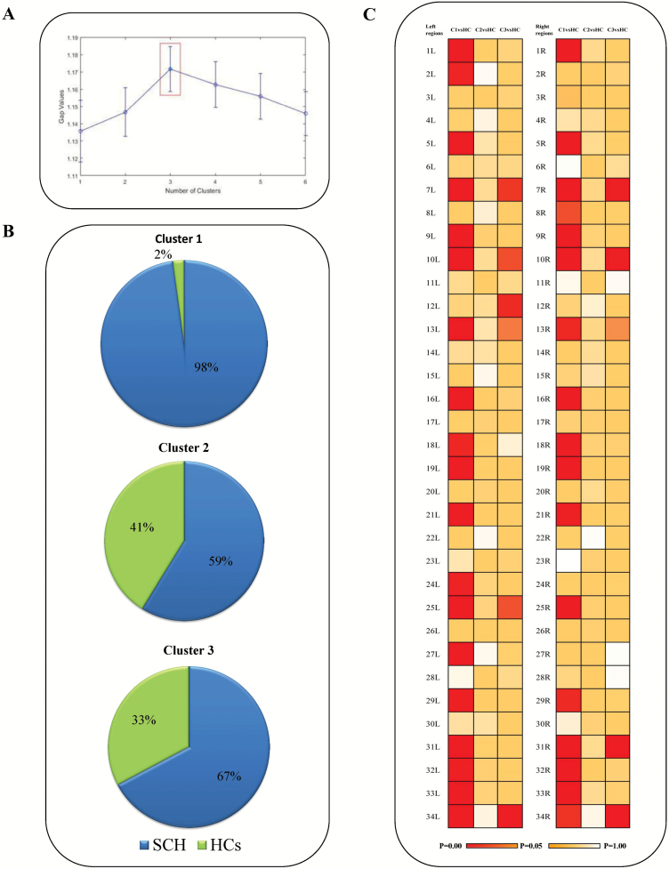
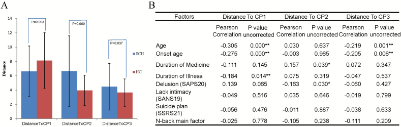
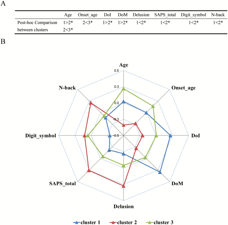
The diagnosis of schizophrenia is thought to embrace several distinct subgroups. The manifold entities in a single clinical patient group increase the variance of biological measures, deflate the group-level estimates of causal factors, and mask the presence of treatment effects. However, reliable neurobiological boundaries to differentiate these subgroups remain elusive. Since cortical thinning is a well-established feature in schizophrenia, we investigated if individuals (patients and healthy controls) with similar patterns of regional cortical thickness form naturally occurring morphological subtypes. K-means algorithm clustering was applied to regional cortical thickness values obtained from 256 structural MRI scans (179 patients with schizophrenia and 77 healthy controls [HCs]). GAP statistics revealed three clusters with distinct regional thickness patterns. The specific patterns of cortical thinning, clinical characteristics, and cognitive function of each clustered subgroup were assessed. The three clusters based on thickness patterns comprised of a morphologically impoverished subgroup (25% patients, 1% HCs), an intermediate subgroup (47% patients, 46% HCs), and an intact subgroup (28% patients, 53% HCs). The differences of clinical features among three clusters pertained to age-of-onset, N-back performance, duration exposure to treatment, total burden of positive symptoms, and severity of delusions. Particularly, the morphologically impoverished group had deficits in N-back performance and less severe positive symptom burden. The data-driven neuroimaging approach illustrates the occurrence of morphologically separable subgroups in schizophrenia, with distinct clinical characteristics. We infer that the anatomical heterogeneity of schizophrenia arises from both pathological deviance and physiological variance. We advocate using MRI-guided stratification for clinical trials as well as case-control investigations in schizophrenia.
精神分裂症的诊断被认为包含几个不同的亚组。单一临床患者群体中的多种实体增加了生物学测量的方差,降低了因果因素的组水平估计,并掩盖了治疗效果的存在。然而,区分这些亚组的可靠神经生物学界限仍然难以捉摸。由于皮质变薄是精神分裂症中一个已确立的特征,我们研究了具有相似区域皮质厚度模式的个体(患者和健康对照)是否形成自然发生的形态学亚型。将K均值算法聚类应用于从256次结构MRI扫描(179例精神分裂症患者和77例健康对照[HCs])获得的区域皮质厚度值。间隙统计揭示了三个具有不同区域厚度模式的聚类。评估了每个聚类亚组的皮质变薄的特定模式、临床特征和认知功能。基于厚度模式的三个聚类包括一个形态学受损亚组(25%的患者,1%的HCs)、一个中间亚组(47%的患者,46%的HCs)和一个完整亚组(28%的患者,53%的HCs)。三个聚类之间临床特征的差异涉及发病年龄、N-back表现、治疗暴露持续时间、阳性症状的总负担和妄想的严重程度。特别是,形态学受损组在N-back表现方面存在缺陷,阳性症状负担较轻。这种数据驱动的神经影像学方法说明了精神分裂症中存在形态学上可分离的亚组,具有不同的临床特征。我们推断,精神分裂症的解剖学异质性源于病理偏差和生理差异。我们主张在精神分裂症的临床试验以及病例对照研究中使用MRI引导的分层。