Bai Siwei, Encke Jörg, Obando-Leitón Miguel, Weiß Robin, Schäfer Friederike, Eberharter Jakob, Böhnke Frank, Hemmert Werner
Department of Electrical and Computer Engineering, Technical University of Munich, Munich, Germany.
Munich School of Bioengineering, Technical University of Munich, Garching, Germany.
Front Neurosci. 2019 Dec 5;13:1312. doi: 10.3389/fnins.2019.01312. eCollection 2019.
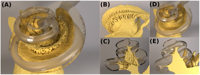
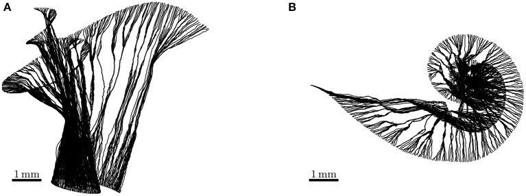
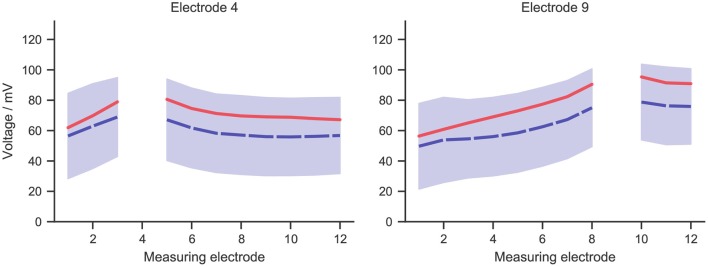
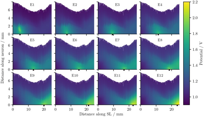
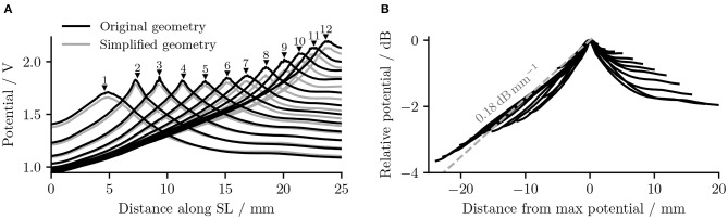
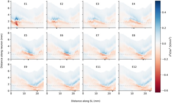
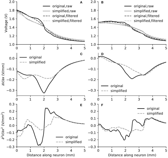
Many detailed features of the cochlear anatomy have not been included in existing 3D cochlear models, including the microstructures inside the modiolar bone, which in turn determines the path of auditory nerve fibers (ANFs). We captured the intricate modiolar microstructures in a 3D human cochlea model reconstructed from μCT scans. A new algorithm was developed to reconstruct ANFs running through the microstructures within the model. Using the finite element method, we calculated the electrical potential as well as its first and second spatial derivatives along each ANF elicited by the cochlear implant electrodes. Simulation results of electrical potential was validated against intracochlear potential measurements. Comparison was then made with a simplified model without the microstructures within the cochlea. When the stimulus was delivered from an electrode located deeper in the apex, the extent of the auditory nerve influenced by a higher electric potential grew larger; at the same time, the maximal potential value at the auditory nerve also became larger. The electric potential decayed at a faster rate toward the base of the cochlea than toward the apex. Compared to the cochlear model incorporating the modiolar microstructures, the simplified version resulted in relatively small differences in electric potential. However, in terms of the first and second derivatives of electric potential along the fibers, which are relevant for the initiation of action potentials, the two models exhibited large differences: maxima in both derivatives with the detailed model were larger by a factor of 1.5 (first derivative) and 2 (second derivative) in the exemplary fibers. More importantly, these maxima occurred at different locations, and opposite signs were found for the values of second derivatives between the two models at parts along the fibers. Hence, while one model predicts depolarization and spike initiation at a given location, the other may instead predict a hyperpolarization. Although a cochlear model with fewer details seems sufficient for analysing the current spread in the cochlear ducts, a detailed-segmented cochlear model is required for the reconstruction of ANF trajectories through the modiolus, as well as the prediction of firing thresholds and spike initiation sites.
现有的三维耳蜗模型并未涵盖耳蜗解剖结构的许多详细特征,包括蜗轴骨内部的微观结构,而蜗轴骨内部的微观结构又决定了听觉神经纤维(ANF)的路径。我们通过μCT扫描重建的三维人体耳蜗模型中捕捉到了复杂的蜗轴微观结构。开发了一种新算法来重建贯穿模型内部微观结构的ANF。使用有限元方法,我们计算了人工耳蜗电极沿每条ANF引发的电势及其一阶和二阶空间导数。电势的模拟结果与耳蜗内电势测量值进行了验证。然后与一个没有耳蜗内部微观结构的简化模型进行了比较。当刺激从位于蜗顶较深处的电极传递时,受较高电势影响的听觉神经范围变大;同时,听觉神经处的最大电势值也变大。电势向耳蜗底部衰减的速度比向蜗顶衰减的速度更快。与包含蜗轴微观结构的耳蜗模型相比,简化版本在电势方面导致的差异相对较小。然而,就沿纤维的电势一阶和二阶导数而言,这与动作电位的引发相关,两个模型表现出很大差异:在示例纤维中,详细模型的两个导数最大值分别比简化模型大1.5倍(一阶导数)和2倍(二阶导数)。更重要的是,这些最大值出现在不同位置,并且在纤维沿线的部分,两个模型的二阶导数的值具有相反的符号。因此,虽然一个模型预测在给定位置会发生去极化和动作电位起始,但另一个模型可能预测会发生超极化。虽然一个细节较少的耳蜗模型似乎足以分析耳蜗管中的电流传播,但需要一个详细分段的耳蜗模型来重建通过蜗轴的ANF轨迹,以及预测放电阈值和动作电位起始位点。