Faculty of Psychology and Neuroscience, Department of Cognitive Neuroscience, Maastricht University, P.O. Box 616, 6200 MD, Maastricht, The Netherlands.
Maastricht Brain Imaging Center (M-BIC), Maastricht University, P.O. Box 616, 6200 MD, Maastricht, The Netherlands.
Commun Biol. 2020 Jan 22;3(1):40. doi: 10.1038/s42003-020-0764-0.
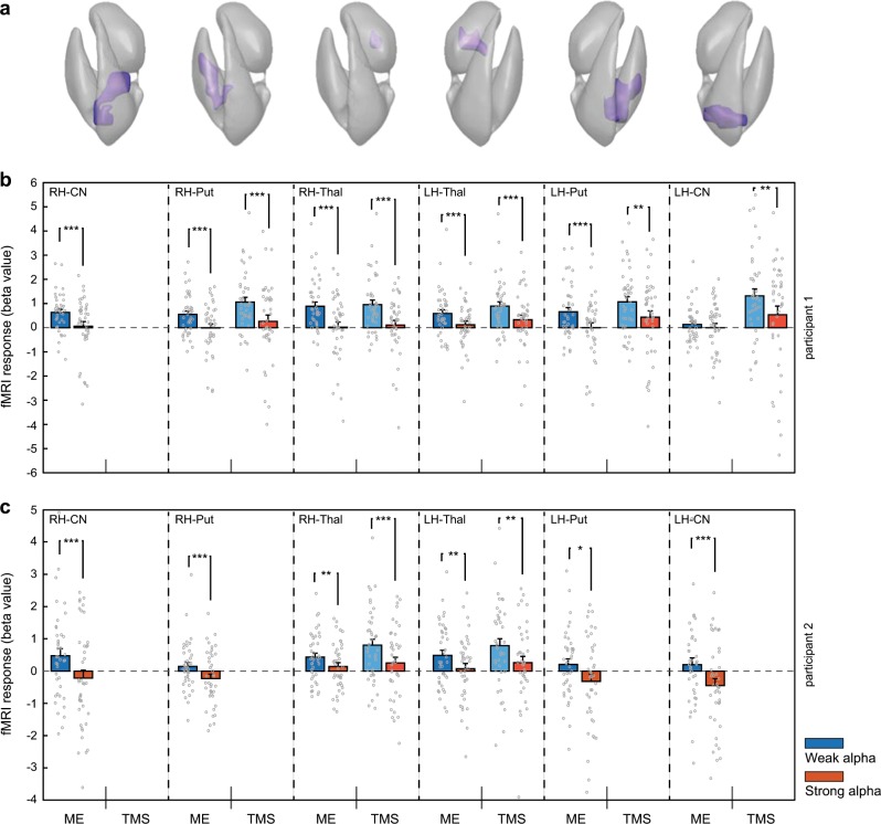
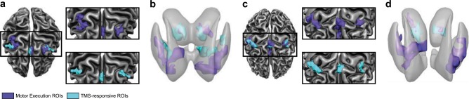
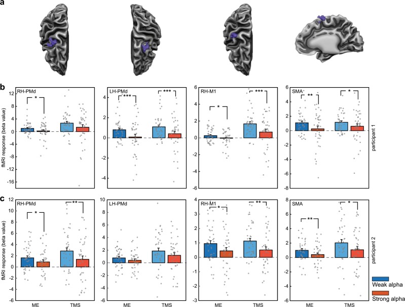
Despite growing interest, the causal mechanisms underlying human neural network dynamics remain elusive. Transcranial Magnetic Stimulation (TMS) allows to noninvasively probe neural excitability, while concurrent fMRI can log the induced activity propagation through connected network nodes. However, this approach ignores ongoing oscillatory fluctuations which strongly affect network excitability and concomitant behavior. Here, we show that concurrent TMS-EEG-fMRI enables precise and direct monitoring of causal dependencies between oscillatory states and signal propagation throughout cortico-subcortical networks. To demonstrate the utility of this multimodal triad, we assessed how pre-TMS EEG power fluctuations influenced motor network activations induced by subthreshold TMS to right dorsal premotor cortex. In participants with adequate motor network reactivity, strong pre-TMS alpha power reduced TMS-evoked hemodynamic activations throughout the bilateral cortico-subcortical motor system (including striatum and thalamus), suggesting shunted network connectivity. Concurrent TMS-EEG-fMRI opens an exciting noninvasive avenue of subject-tailored network research into dynamic cognitive circuits and their dysfunction.
尽管人们越来越感兴趣,但人类神经网络动力学的因果机制仍然难以捉摸。经颅磁刺激 (TMS) 允许非侵入性地探测神经兴奋性,而同时进行的 fMRI 可以记录通过连接的网络节点诱导的活动传播。然而,这种方法忽略了正在进行的振荡波动,这些波动强烈影响网络兴奋性和伴随的行为。在这里,我们表明,同时进行的 TMS-EEG-fMRI 能够精确和直接监测振荡状态之间的因果关系以及皮质下网络中的信号传播。为了展示这种多模态三联体的实用性,我们评估了 TMS 前 EEG 功率波动如何影响由右侧背侧运动前皮质的亚阈 TMS 引起的运动网络激活。在具有足够运动网络反应性的参与者中,强 TMS 前 alpha 功率降低了双侧皮质下运动系统(包括纹状体和丘脑)中 TMS 诱发的血液动力学激活,表明网络连接分流。同时进行的 TMS-EEG-fMRI 为针对动态认知电路及其功能障碍的个体化网络研究开辟了一条令人兴奋的非侵入性途径。