International Iberian Nanotechnology Laboratory, Avenida Mestre José Veiga s/n, Braga, Portugal.
Department of Applied Physics, University of Santiago de Compostela, E-15782, Santiago de Compostela, Spain.
Sci Rep. 2020 Jan 24;10(1):1122. doi: 10.1038/s41598-020-57885-z.
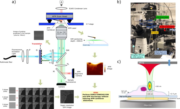
Correlating data from different microscopy techniques holds the potential to discover new facets of signaling events in cellular biology. Here we report for the first time a hardware set-up capable of achieving simultaneous co-localized imaging of spatially correlated far-field super-resolution fluorescence microscopy and atomic force microscopy, a feat only obtained until now by fluorescence microscopy set-ups with spatial resolution restricted by the Abbe diffraction limit. We detail system integration and demonstrate system performance using sub-resolution fluorescent beads and applied to a test sample consisting of human bone osteosarcoma epithelial cells, with plasma membrane transporter 1 (MCT1) tagged with an enhanced green fluorescent protein (EGFP) at the N-terminal.
从不同的显微镜技术中获取相关数据有可能揭示细胞生物学中信号事件的新方面。在这里,我们首次报告了一种硬件设置,能够实现空间相关的远场超分辨率荧光显微镜和原子力显微镜的同时共定位成像,这是迄今为止仅通过空间分辨率受阿贝衍射极限限制的荧光显微镜设置才能实现的壮举。我们详细介绍了系统集成,并使用亚分辨率荧光珠演示了系统性能,然后将其应用于包含人骨肉瘤上皮细胞的测试样本,该样本的细胞膜转运蛋白 1(MCT1)在 N 端标记有增强型绿色荧光蛋白(EGFP)。