Malinauskaite Ieva, Hofmeister Jeremy, Burgermeister Simon, Neroladaki Angeliki, Hamard Marion, Montet Xavier, Boudabbous Sana
Geneva University Hospital, Diagnosis Department, Radiology Division, Rue Gabrielle-Perret-Gentil 4, 1211 Geneva 4, Switzerland.
Sarcoma. 2020 Jan 7;2020:7163453. doi: 10.1155/2020/7163453. eCollection 2020.
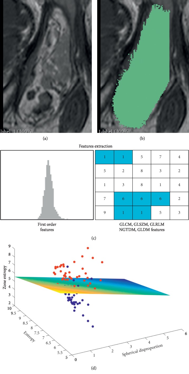
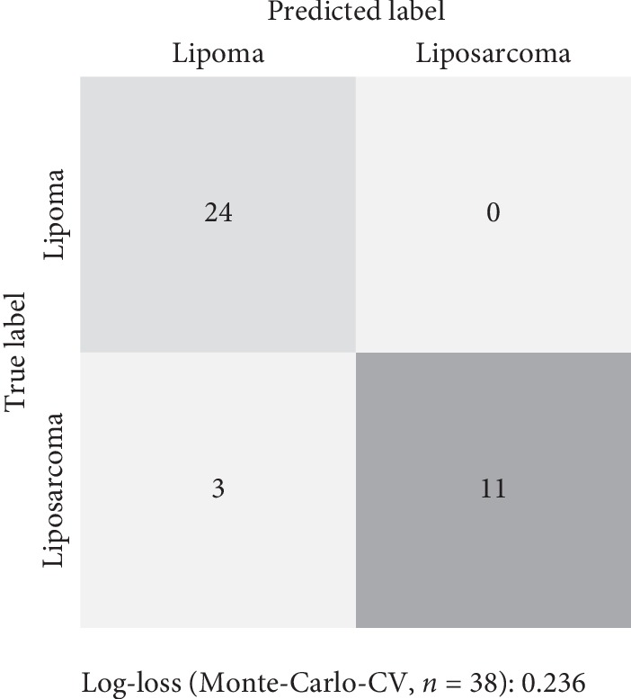
Distinguishing lipoma from liposarcoma is challenging on conventional MRI examination. In case of uncertain diagnosis following MRI, further invasive procedure (percutaneous biopsy or surgery) is often required to allow for diagnosis based on histopathological examination. Radiomics and machine learning allow for several types of pathologies encountered on radiological images to be automatically and reliably distinguished. The aim of the study was to assess the contribution of radiomics and machine learning in the differentiation between soft-tissue lipoma and liposarcoma on preoperative MRI and to assess the diagnostic accuracy of a machine-learning model compared to musculoskeletal radiologists. 86 radiomics features were retrospectively extracted from volume-of-interest on T1-weighted spin-echo 1.5 and 3.0 Tesla MRI of 38 soft-tissue tumors (24 lipomas and 14 liposarcomas, based on histopathological diagnosis). These radiomics features were then used to train a machine-learning classifier to distinguish lipoma and liposarcoma. The generalization performance of the machine-learning model was assessed using Monte-Carlo cross-validation and receiver operating characteristic curve analysis (ROC-AUC). Finally, the performance of the machine-learning model was compared to the accuracy of three specialized musculoskeletal radiologists using the McNemar test. Machine-learning classifier accurately distinguished lipoma and liposarcoma, with a ROC-AUC of 0.926. Notably, it performed better than the three specialized musculoskeletal radiologists reviewing the same patients, who achieved ROC-AUC of 0.685, 0.805, and 0.785. Despite being developed on few cases, the trained machine-learning classifier accurately distinguishes lipoma and liposarcoma on preoperative MRI, with better performance than specialized musculoskeletal radiologists.
在传统的MRI检查中,区分脂肪瘤和脂肪肉瘤具有挑战性。如果MRI检查后诊断不确定,通常需要进一步的侵入性检查(经皮活检或手术),以便基于组织病理学检查进行诊断。放射组学和机器学习能够自动且可靠地区分放射图像上出现的几种病理类型。本研究的目的是评估放射组学和机器学习在术前MRI上区分软组织脂肪瘤和脂肪肉瘤中的作用,并评估与肌肉骨骼放射科医生相比,机器学习模型的诊断准确性。回顾性地从38例软组织肿瘤(根据组织病理学诊断,其中24例为脂肪瘤,14例为脂肪肉瘤)的T1加权自旋回波1.5和3.0特斯拉MRI的感兴趣体积中提取了86个放射组学特征。然后,这些放射组学特征被用于训练一个机器学习分类器,以区分脂肪瘤和脂肪肉瘤。使用蒙特卡洛交叉验证和受试者工作特征曲线分析(ROC-AUC)评估机器学习模型的泛化性能。最后,使用McNemar检验将机器学习模型的性能与三位专业肌肉骨骼放射科医生的准确性进行比较。机器学习分类器能够准确区分脂肪瘤和脂肪肉瘤,ROC-AUC为0.926。值得注意的是,它的表现优于三位评估相同患者的专业肌肉骨骼放射科医生,他们的ROC-AUC分别为0.685、0.805和0.785。尽管是基于少数病例开发的,但经过训练 的机器学习分类器在术前MRI上能够准确区分脂肪瘤和脂肪肉瘤,其性能优于专业肌肉骨骼放射科医生。