IRCCS Istituto Ortopedico Galeazzi, Milan, Italy.
Dipartimento di Scienze Biomediche per la Salute, Università degli Studi di Milano, Milan, Italy.
Radiol Med. 2023 Aug;128(8):989-998. doi: 10.1007/s11547-023-01657-y. Epub 2023 Jun 19.
To determine diagnostic performance of MRI radiomics-based machine learning for classification of deep-seated lipoma and atypical lipomatous tumor (ALT) of the extremities.
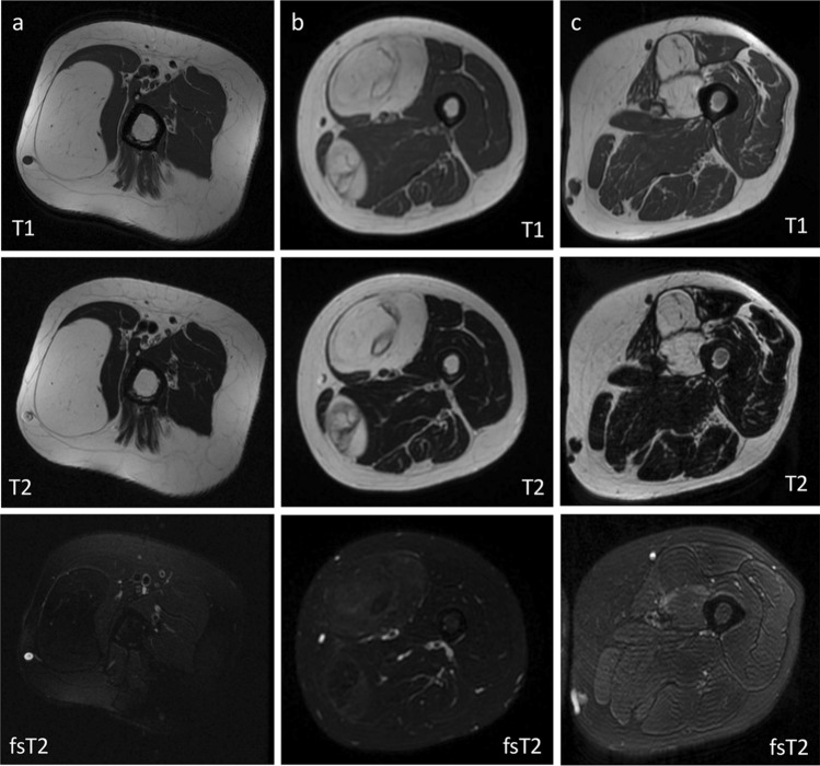
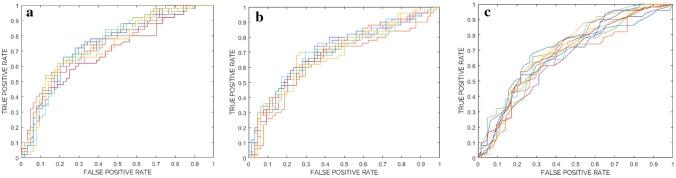
This retrospective study was performed at three tertiary sarcoma centers and included 150 patients with surgically treated and histology-proven lesions. The training-validation cohort consisted of 114 patients from centers 1 and 2 (n = 64 lipoma, n = 50 ALT). The external test cohort consisted of 36 patients from center 3 (n = 24 lipoma, n = 12 ALT). 3D segmentation was manually performed on T1- and T2-weighted MRI. After extraction and selection of radiomic features, three machine learning classifiers were trained and validated using nested fivefold cross-validation. The best-performing classifier according to previous analysis was evaluated and compared to an experienced musculoskeletal radiologist in the external test cohort.
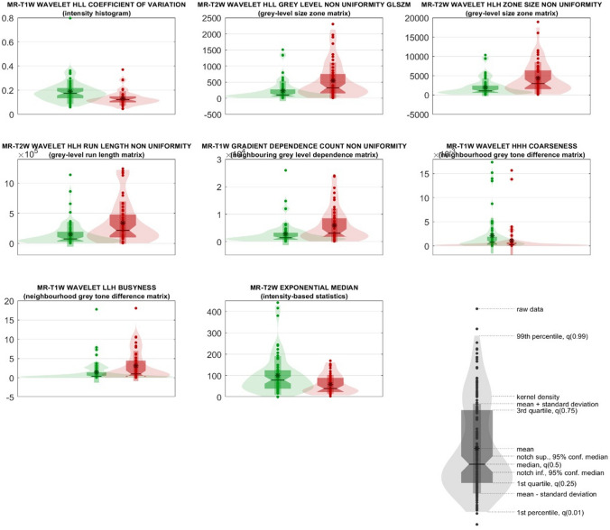
Eight features passed feature selection and were incorporated into the machine learning models. After training and validation (74% ROC-AUC), the best-performing classifier (Random Forest) showed 92% sensitivity and 33% specificity in the external test cohort with no statistical difference compared to the radiologist (p = 0.474).
MRI radiomics-based machine learning may classify deep-seated lipoma and ALT of the extremities with high sensitivity and negative predictive value, thus potentially serving as a non-invasive screening tool to reduce unnecessary referral to tertiary tumor centers.
确定基于 MRI 放射组学的机器学习对四肢深部脂肪瘤和非典型性脂肪肉瘤(ALT)分类的诊断性能。
本回顾性研究在三个三级肉瘤中心进行,纳入了 150 名接受手术治疗和组织学证实病变的患者。训练-验证队列由来自中心 1 和 2 的 114 名患者组成(n=64 例脂肪瘤,n=50 例 ALT)。外部测试队列由来自中心 3 的 36 名患者组成(n=24 例脂肪瘤,n=12 例 ALT)。手动在 T1 和 T2 加权 MRI 上进行 3D 分割。在提取和选择放射组学特征后,使用嵌套五折交叉验证训练和验证了三个机器学习分类器。根据之前的分析,评估并比较最佳表现分类器与外部测试队列中的经验丰富的肌肉骨骼放射科医生。
经过特征选择,有 8 个特征被纳入机器学习模型。经过训练和验证(74%ROC-AUC),表现最佳的分类器(随机森林)在外部测试队列中的敏感性为 92%,特异性为 33%,与放射科医生无统计学差异(p=0.474)。
基于 MRI 放射组学的机器学习可能具有高敏感性和阴性预测值,可以对四肢深部脂肪瘤和 ALT 进行分类,从而有可能成为一种非侵入性的筛选工具,以减少对三级肿瘤中心的不必要转诊。