• 文献检索
  • 文档翻译
  • 深度研究
  • 学术资讯
  • Suppr Zotero 插件Zotero 插件
  • 邀请有礼
  • 套餐&价格
  • 历史记录
应用&插件
Suppr Zotero 插件Zotero 插件浏览器插件Mac 客户端Windows 客户端微信小程序
定价
高级版会员购买积分包购买API积分包
服务
文献检索文档翻译深度研究API 文档MCP 服务
关于我们
关于 Suppr公司介绍联系我们用户协议隐私条款
关注我们

Suppr 超能文献

核心技术专利:CN118964589B侵权必究
粤ICP备2023148730 号-1Suppr @ 2026

文献检索

告别复杂PubMed语法,用中文像聊天一样搜索,搜遍4000万医学文献。AI智能推荐,让科研检索更轻松。

立即免费搜索

文件翻译

保留排版,准确专业,支持PDF/Word/PPT等文件格式,支持 12+语言互译。

免费翻译文档

深度研究

AI帮你快速写综述,25分钟生成高质量综述,智能提取关键信息,辅助科研写作。

立即免费体验

磁粒子成像显示和定量检测磁性纳米颗粒的细胞摄取。

Cellular uptake of magnetic nanoparticles imaged and quantified by magnetic particle imaging.

机构信息

Physikalisch-Technische Bundesanstalt, Berlin, Germany.

Charité - Universitätsmedizin Berlin, corporate member of Freie Universität Berlin, Humboldt-Universität zu Berlin, Berlin Institute of Health, Medizinische Klinik für Kardiologie und Angiologie, Campus Mitte, Berlin, Germany.

出版信息

Sci Rep. 2020 Feb 5;10(1):1922. doi: 10.1038/s41598-020-58853-3.

DOI:10.1038/s41598-020-58853-3
PMID:32024926
原文链接:https://pmc.ncbi.nlm.nih.gov/articles/PMC7002802/
Abstract

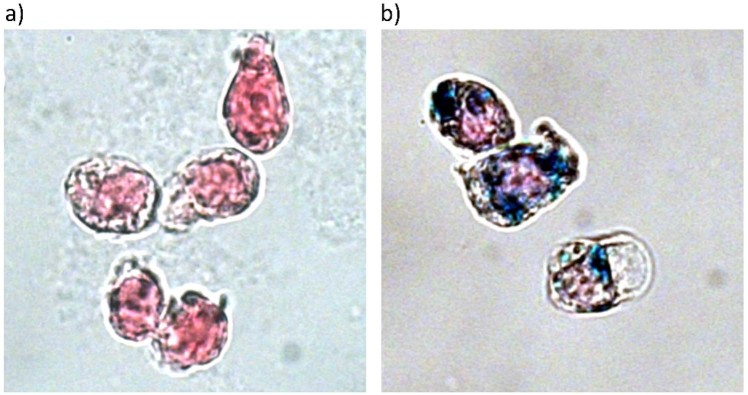
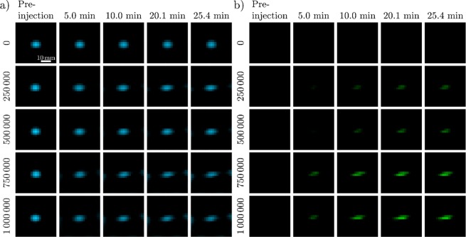
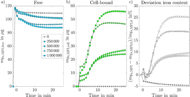
Magnetic particle imaging (MPI) is a non-invasive, non-ionizing imaging technique for the visualization and quantification of magnetic nanoparticles (MNPs). The technique is especially suitable for cell imaging as it offers zero background contribution from the surrounding tissue, high sensitivity, and good spatial and temporal resolutions. Previous studies have demonstrated that the dynamic magnetic behaviour of MNPs changes during cellular binding and internalization. In this study, we demonstrate how this information is encoded in the MPI imaging signal. Through MPI imaging we are able to discriminate between free and cell-bound MNPs in reconstructed images. This technique was used to image and quantify the changes that occur in-vitro when free MNPs come into contact with cells and undergo cellular-uptake over time. The quantitative MPI results were verified by colorimetric measurements of the iron content. The results showed a mean relative difference between the MPI results and the reference method of 23.8% for the quantification of cell-bound MNPs. With this technique, the uptake of MNPs in cells can be imaged and quantified directly from the first MNP cell contact, providing information on the dynamics of cellular uptake.

摘要

磁粒子成像(MPI)是一种用于可视化和量化磁性纳米粒子(MNPs)的非侵入性、非电离成像技术。该技术特别适用于细胞成像,因为它提供了来自周围组织的零背景贡献、高灵敏度以及良好的空间和时间分辨率。先前的研究表明,MNPs 的动态磁行为在细胞结合和内化过程中发生变化。在本研究中,我们展示了如何在 MPI 成像信号中对这些信息进行编码。通过 MPI 成像,我们能够在重建图像中区分游离和细胞结合的 MNPs。这项技术用于成像和量化体外实验中游离 MNPs 与细胞接触并随时间发生细胞摄取时的变化。通过比色法测量铁含量对定量 MPI 结果进行了验证。结果表明,MPI 结果与参考方法定量细胞结合 MNPs 的平均相对差异为 23.8%。通过这项技术,可以直接从第一个 MNP 与细胞接触开始对细胞内的 MNPs 摄取进行成像和定量,提供有关细胞摄取动力学的信息。

https://cdn.ncbi.nlm.nih.gov/pmc/blobs/f373/7002802/5fc423d54fbb/41598_2020_58853_Fig4_HTML.jpg
https://cdn.ncbi.nlm.nih.gov/pmc/blobs/f373/7002802/f186c8e9ba72/41598_2020_58853_Fig1_HTML.jpg
https://cdn.ncbi.nlm.nih.gov/pmc/blobs/f373/7002802/d63928a50f05/41598_2020_58853_Fig2_HTML.jpg
https://cdn.ncbi.nlm.nih.gov/pmc/blobs/f373/7002802/cae61aa5dba2/41598_2020_58853_Fig3_HTML.jpg
https://cdn.ncbi.nlm.nih.gov/pmc/blobs/f373/7002802/5fc423d54fbb/41598_2020_58853_Fig4_HTML.jpg

相似文献

1
Cellular uptake of magnetic nanoparticles imaged and quantified by magnetic particle imaging.磁粒子成像显示和定量检测磁性纳米颗粒的细胞摄取。
Sci Rep. 2020 Feb 5;10(1):1922. doi: 10.1038/s41598-020-58853-3.
2
Cell Tracking by Magnetic Particle Imaging: Methodology for Labeling THP-1 Monocytes with Magnetic Nanoparticles for Cellular Imaging.细胞示踪的磁粒子成像:用磁性纳米颗粒标记 THP-1 单核细胞进行细胞成像的方法。
Cells. 2022 Sep 16;11(18):2892. doi: 10.3390/cells11182892.
3
Magnetic Nanoparticles in Macrophages and Cancer Cells Exhibit Different Signal Behavior on Magnetic Particle Imaging.磁纳米粒子在巨噬细胞和癌细胞中的磁粒子成像表现出不同的信号行为。
J Nanosci Nanotechnol. 2019 Nov 1;19(11):6857-6865. doi: 10.1166/jnn.2019.16619.
4
Characterization of magnetic nanoparticle systems with respect to their magnetic particle imaging performance.关于磁性纳米颗粒系统的磁粒子成像性能的表征
Biomed Tech (Berl). 2013 Dec;58(6):535-45. doi: 10.1515/bmt-2013-0013.
5
Bimodal intravascular volumetric imaging combining OCT and MPI.结合 OCT 和 MPI 的双模态血管内容积成像。
Med Phys. 2019 Mar;46(3):1371-1383. doi: 10.1002/mp.13388. Epub 2019 Feb 14.
6
Counting cells in motion by quantitative real-time magnetic particle imaging.通过定量实时磁粒子成像技术对运动中的细胞进行计数。
Sci Rep. 2024 Feb 21;14(1):4253. doi: 10.1038/s41598-024-54784-5.
7
Experimental and simulation studies on the behavior of signal harmonics in magnetic particle imaging.磁颗粒成像中信号谐波行为的实验与模拟研究
Radiol Phys Technol. 2013 Jul;6(2):399-414. doi: 10.1007/s12194-013-0213-6. Epub 2013 Apr 16.
8
Magnetorelaxometry procedures for quantitative imaging and characterization of magnetic nanoparticles in biomedical applications.用于生物医学应用中磁性纳米颗粒定量成像和表征的磁弛豫测量方法。
Biomed Tech (Berl). 2015 Oct;60(5):427-43. doi: 10.1515/bmt-2015-0055.
9
Seeing SPIOs Directly In Vivo with Magnetic Particle Imaging.利用磁粒子成像在体内直接观察超顺磁性氧化铁纳米颗粒
Mol Imaging Biol. 2017 Jun;19(3):385-390. doi: 10.1007/s11307-017-1081-y.
10
Optimizing magnetite nanoparticles for mass sensitivity in magnetic particle imaging.优化磁铁矿纳米颗粒以提高磁粒子成像的质量灵敏度。
Med Phys. 2011 Mar;38(3):1619-26. doi: 10.1118/1.3554646.

引用本文的文献

1
The Role of Glycocalyx Diversity and Thickness for Nanoparticle Internalization in M1-/M2-like Macrophages.糖萼多样性和厚度在M1型/M2型巨噬细胞中对纳米颗粒内化的作用
Nano Lett. 2024 Dec 11;24(49):15607-15614. doi: 10.1021/acs.nanolett.4c04004. Epub 2024 Dec 2.
2
Rapid cellular uptake of citrate-coated iron oxide nanoparticles unaffected by cell-surface glycosaminoglycans.柠檬酸盐包被的氧化铁纳米颗粒可被细胞快速摄取,且不受细胞表面糖胺聚糖的影响。
Nanoscale Adv. 2024 Jun 13;6(15):3825-3837. doi: 10.1039/d4na00277f. eCollection 2024 Jul 23.
3
Surfactant-driven optimization of iron-based nanoparticle synthesis: a study on magnetic hyperthermia and endothelial cell uptake.

本文引用的文献

1
Correction of linear system drifts in magnetic particle imaging.磁粒子成像中线性系统漂移的校正。
Phys Med Biol. 2019 Jun 20;64(12):125013. doi: 10.1088/1361-6560/ab2480.
2
Intracellular dynamics of superparamagnetic iron oxide nanoparticles for magnetic particle imaging.超顺磁性氧化铁纳米颗粒的细胞内动力学用于磁粒子成像。
Nanoscale. 2019 Apr 23;11(16):7771-7780. doi: 10.1039/c9nr01395d.
3
Very small superparamagnetic iron oxide nanoparticles: Long-term fate and metabolic processing in atherosclerotic mice.非常小的超顺磁性氧化铁纳米颗粒:在动脉粥样硬化小鼠中的长期归宿和代谢过程。
表面活性剂驱动的铁基纳米颗粒合成优化:磁热疗与内皮细胞摄取研究
Nanoscale Adv. 2023 Oct 4;5(21):5859-5869. doi: 10.1039/d3na00540b. eCollection 2023 Oct 24.
4
In vivo tracking of adenoviral-transduced iron oxide-labeled bone marrow-derived dendritic cells using magnetic particle imaging.体内示踪腺病毒转铁蛋白标记的骨髓来源树突状细胞的磁粒子成像研究
Eur Radiol Exp. 2023 Aug 15;7(1):42. doi: 10.1186/s41747-023-00359-4.
5
3D modeling of in vivo MRI-guided nano-photothermal therapy mediated by magneto-plasmonic nanohybrids.基于磁等离子体纳米杂化的体内 MRI 引导的纳米光热治疗的 3D 建模。
Biomed Eng Online. 2023 Aug 1;22(1):77. doi: 10.1186/s12938-023-01131-w.
6
Monitoring magnetic nanoparticle clustering and immobilization with thermal noise magnetometry using optically pumped magnetometers.使用光泵磁力计通过热噪声磁力测量法监测磁性纳米颗粒的聚集和固定化。
Nanoscale Adv. 2023 Mar 15;5(8):2341-2351. doi: 10.1039/d3na00016h. eCollection 2023 Apr 11.
7
Dual Magnetic Particle Imaging and Akaluc Bioluminescence Imaging for Tracking Cancer Cell Metastasis.双顺磁纳米粒子成像和 Akaluc 生物发光成像用于追踪癌细胞转移。
Tomography. 2023 Jan 25;9(1):178-194. doi: 10.3390/tomography9010016.
8
Cell Tracking by Magnetic Particle Imaging: Methodology for Labeling THP-1 Monocytes with Magnetic Nanoparticles for Cellular Imaging.细胞示踪的磁粒子成像:用磁性纳米颗粒标记 THP-1 单核细胞进行细胞成像的方法。
Cells. 2022 Sep 16;11(18):2892. doi: 10.3390/cells11182892.
9
Kinetics of nanoparticle uptake into and distribution in human cells.纳米颗粒被人体细胞摄取及其在细胞内分布的动力学
Nanoscale Adv. 2021 Mar 18;3(8):2196-2212. doi: 10.1039/d0na00716a. eCollection 2021 Apr 20.
10
Long-Term Clearance and Biodistribution of Magnetic Nanoparticles Assessed by AC Biosusceptometry.通过交流生物磁测量法评估磁性纳米颗粒的长期清除率和生物分布
Materials (Basel). 2022 Mar 14;15(6):2121. doi: 10.3390/ma15062121.
Nanomedicine. 2018 Nov;14(8):2575-2586. doi: 10.1016/j.nano.2018.07.013. Epub 2018 Sep 1.
4
In vivo tracking and quantification of inhaled aerosol using magnetic particle imaging towards inhaled therapeutic monitoring.利用磁粒子成像技术对吸入性气溶胶进行体内跟踪和定量,实现吸入性治疗监测。
Theranostics. 2018 Jun 8;8(13):3676-3687. doi: 10.7150/thno.26608. eCollection 2018.
5
Improved sensitivity and limit-of-detection using a receive-only coil in magnetic particle imaging.使用仅接收线圈的磁共振成像技术提高灵敏度和检测极限。
Phys Med Biol. 2018 Jul 2;63(13):13NT02. doi: 10.1088/1361-6560/aacb87.
6
Joint Reconstruction of Tracer Distribution and Background in Magnetic Particle Imaging.磁共振粒子成像中示踪剂分布与本底的联合重建
IEEE Trans Med Imaging. 2018 May;37(5):1192-1203. doi: 10.1109/TMI.2017.2777878.
7
Tomographic magnetic particle imaging of cancer targeted nanoparticles.癌症靶向纳米粒子的层析磁共振粒子成像。
Nanoscale. 2017 Dec 7;9(47):18723-18730. doi: 10.1039/c7nr05502a.
8
Towards Picogram Detection of Superparamagnetic Iron-Oxide Particles Using a Gradiometric Receive Coil.利用梯度接收线圈实现对超顺磁性氧化铁粒子的皮克级检测。
Sci Rep. 2017 Jul 31;7(1):6872. doi: 10.1038/s41598-017-06992-5.
9
Magnetic Particle Imaging: A Novel in Vivo Imaging Platform for Cancer Detection.磁性粒子成像:一种用于癌症检测的新型活体成像平台。
Nano Lett. 2017 Mar 8;17(3):1648-1654. doi: 10.1021/acs.nanolett.6b04865. Epub 2017 Feb 21.
10
Magnetic Particle Imaging for High Temporal Resolution Assessment of Aneurysm Hemodynamics.用于动脉瘤血流动力学高时间分辨率评估的磁粒子成像
PLoS One. 2016 Aug 5;11(8):e0160097. doi: 10.1371/journal.pone.0160097. eCollection 2016.