• 文献检索
  • 文档翻译
  • 深度研究
  • 学术资讯
  • Suppr Zotero 插件Zotero 插件
  • 邀请有礼
  • 套餐&价格
  • 历史记录
应用&插件
Suppr Zotero 插件Zotero 插件浏览器插件Mac 客户端Windows 客户端微信小程序
定价
高级版会员购买积分包购买API积分包
服务
文献检索文档翻译深度研究API 文档MCP 服务
关于我们
关于 Suppr公司介绍联系我们用户协议隐私条款
关注我们

Suppr 超能文献

核心技术专利:CN118964589B侵权必究
粤ICP备2023148730 号-1Suppr @ 2026

文献检索

告别复杂PubMed语法,用中文像聊天一样搜索,搜遍4000万医学文献。AI智能推荐,让科研检索更轻松。

立即免费搜索

文件翻译

保留排版,准确专业,支持PDF/Word/PPT等文件格式,支持 12+语言互译。

免费翻译文档

深度研究

AI帮你快速写综述,25分钟生成高质量综述,智能提取关键信息,辅助科研写作。

立即免费体验

HPF1 完成了 PARP 活性部位,以促进 DNA 损伤诱导的 ADP-核糖基化。

HPF1 completes the PARP active site for DNA damage-induced ADP-ribosylation.

机构信息

Sir William Dunn School of Pathology, University of Oxford, Oxford, UK.

MRC Laboratory of Molecular Biology, Cambridge, UK.

出版信息

Nature. 2020 Mar;579(7800):598-602. doi: 10.1038/s41586-020-2013-6. Epub 2020 Feb 6.

DOI:10.1038/s41586-020-2013-6
PMID:32028527
原文链接:https://pmc.ncbi.nlm.nih.gov/articles/PMC7104379/
Abstract

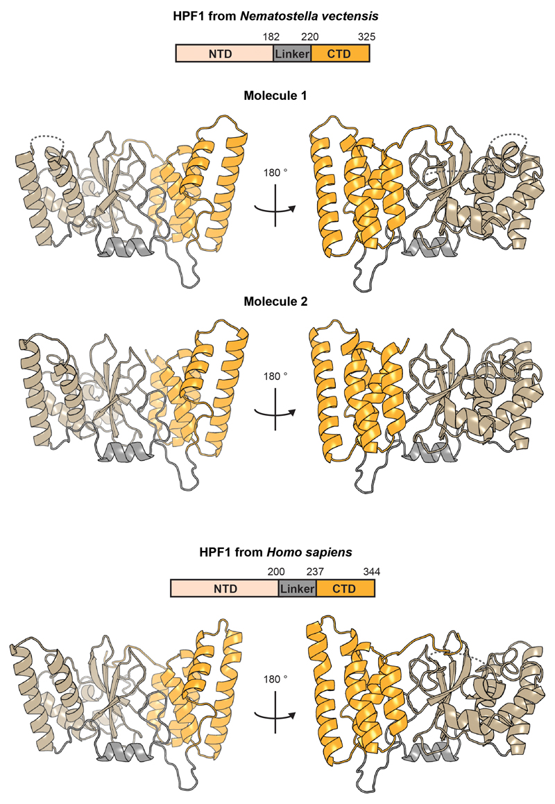
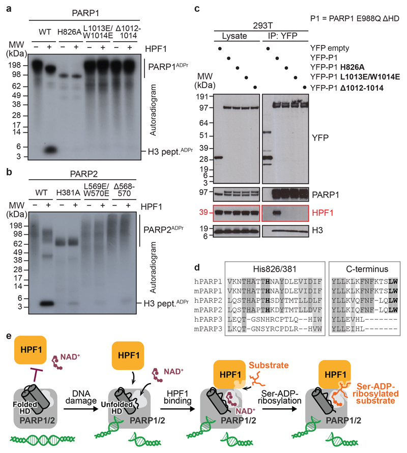
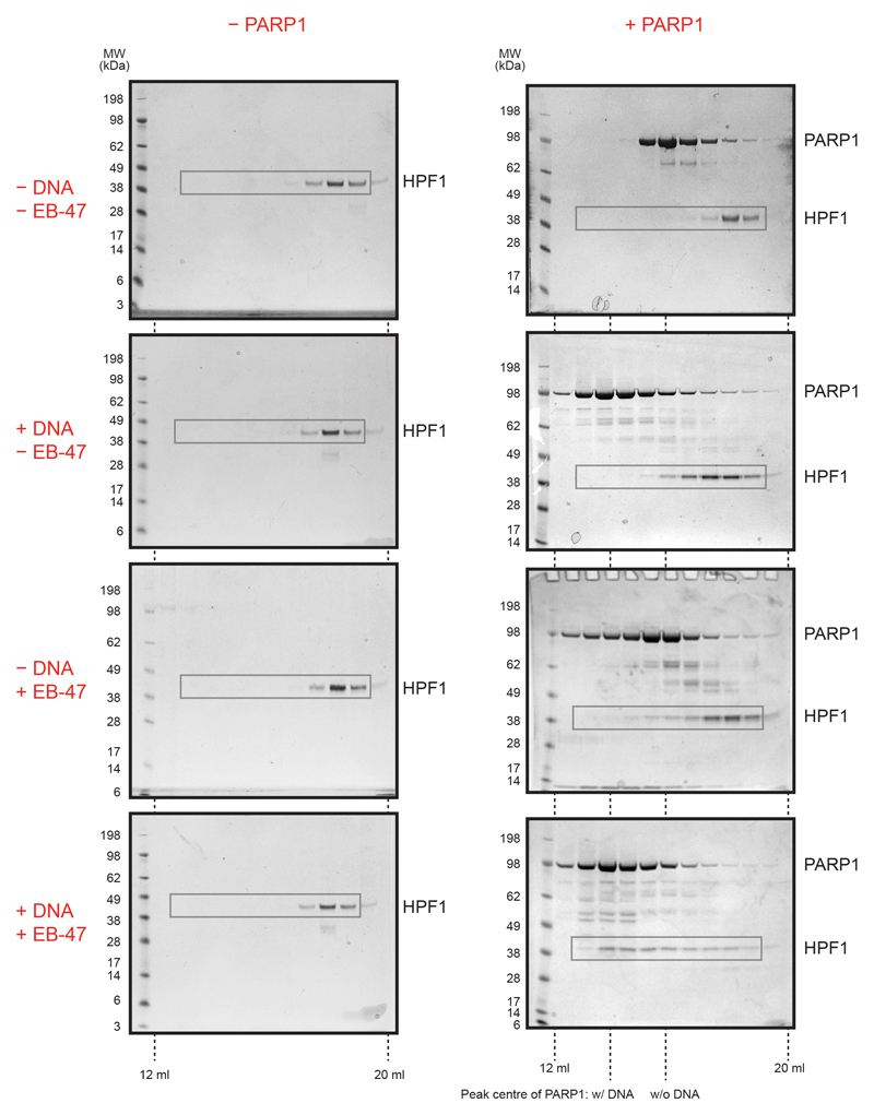
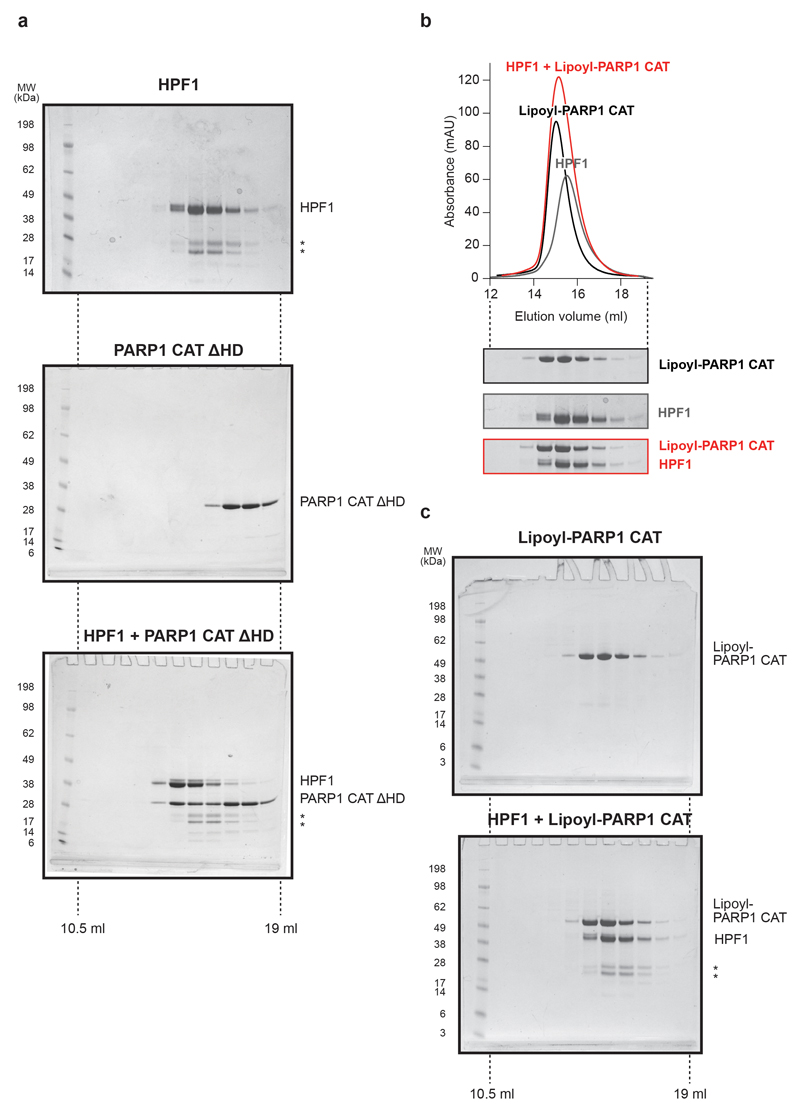
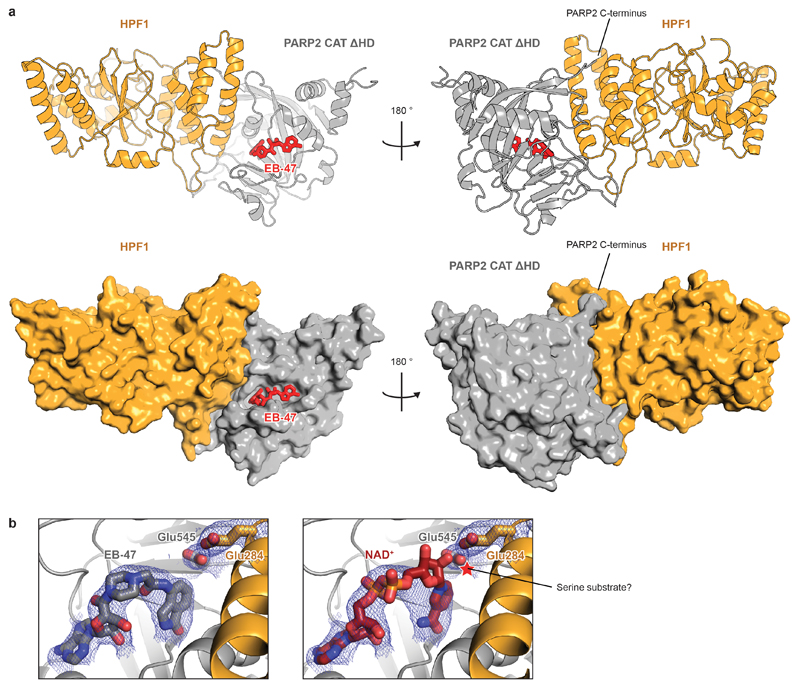
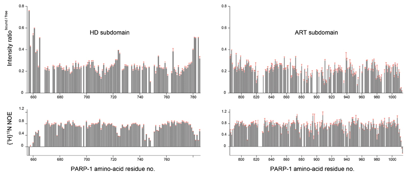
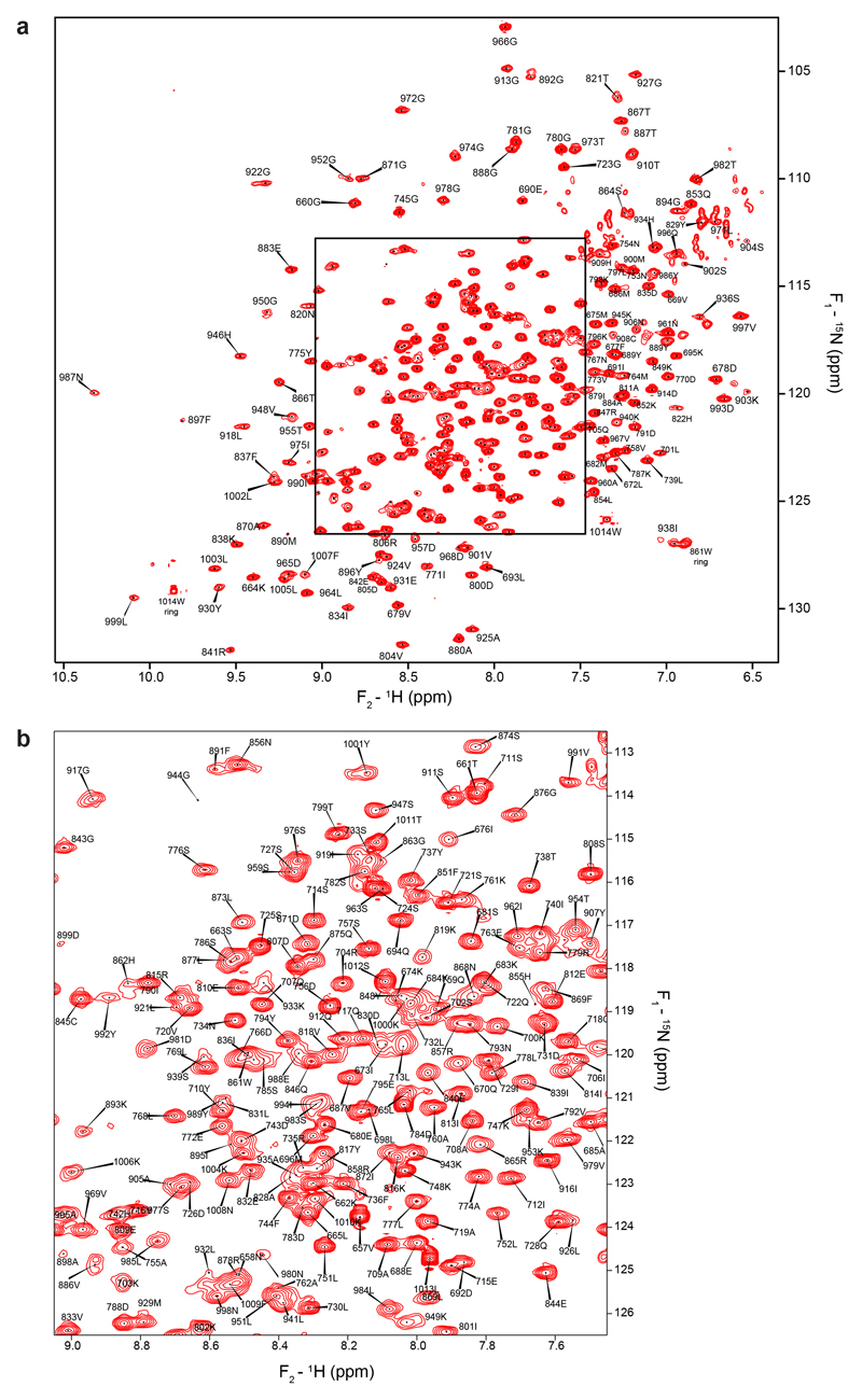
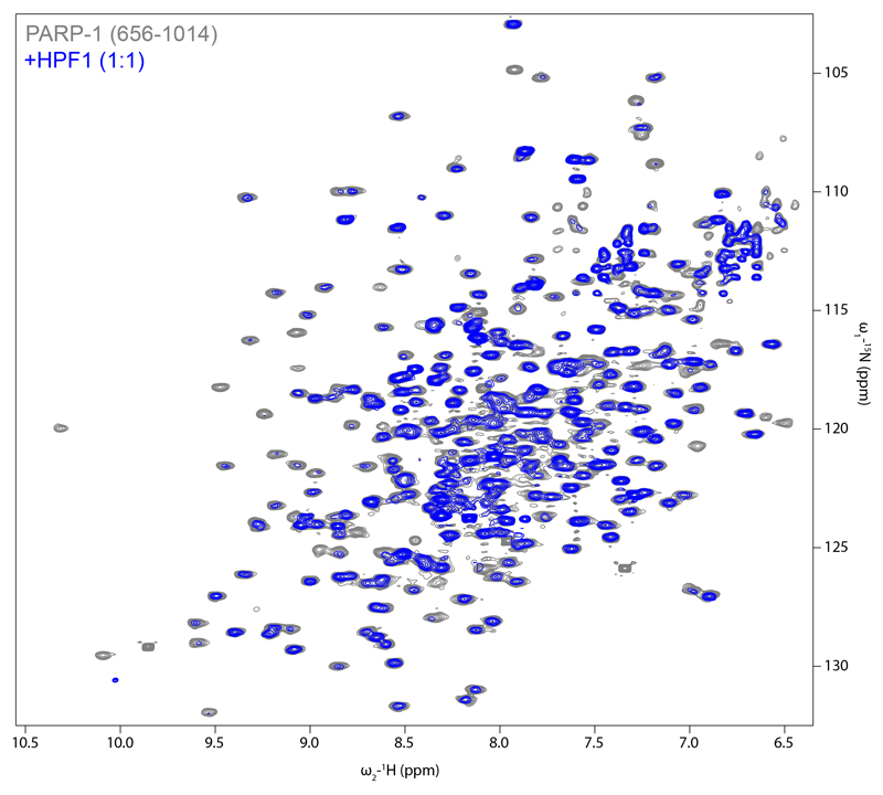
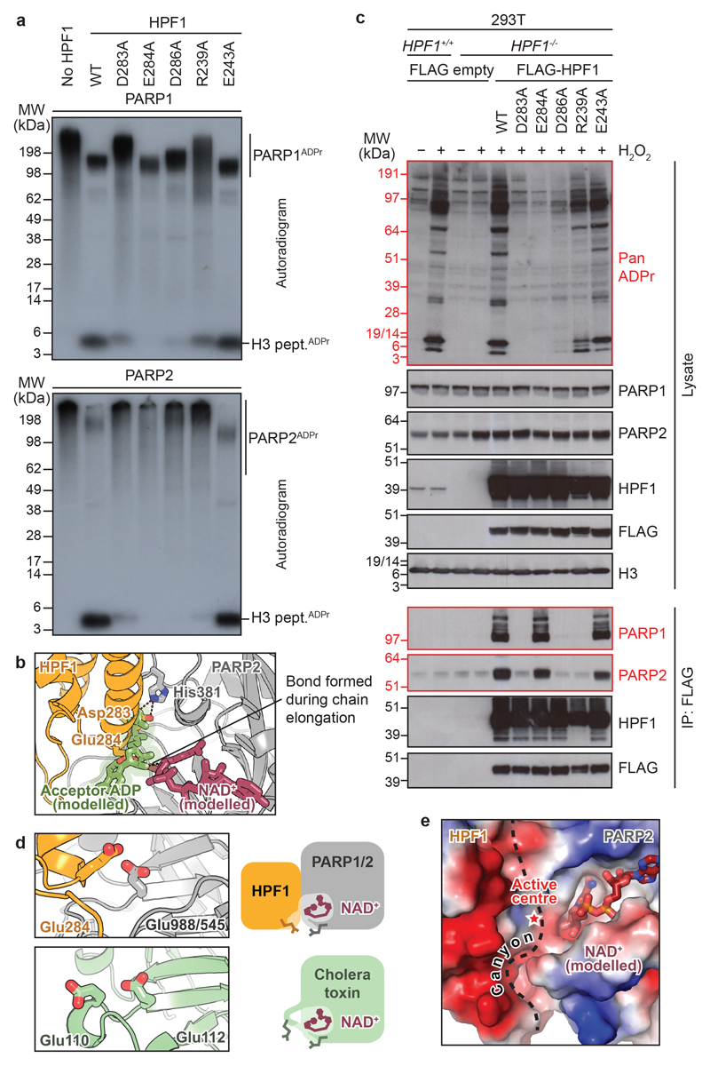
The anti-cancer drug target poly(ADP-ribose) polymerase 1 (PARP1) and its close homologue, PARP2, are early responders to DNA damage in human cells. After binding to genomic lesions, these enzymes use NAD to modify numerous proteins with mono- and poly(ADP-ribose) signals that are important for the subsequent decompaction of chromatin and the recruitment of repair factors. These post-translational modifications are predominantly serine-linked and require the accessory factor HPF1, which is specific for the DNA damage response and switches the amino acid specificity of PARP1 and PARP2 from aspartate or glutamate to serine residues. Here we report a co-structure of HPF1 bound to the catalytic domain of PARP2 that, in combination with NMR and biochemical data, reveals a composite active site formed by residues from HPF1 and PARP1 or PARP2 . The assembly of this catalytic centre is essential for the addition of ADP-ribose moieties after DNA damage in human cells. In response to DNA damage and occupancy of the NAD-binding site, the interaction of HPF1 with PARP1 or PARP2 is enhanced by allosteric networks that operate within the PARP proteins, providing an additional level of regulation in the induction of the DNA damage response. As HPF1 forms a joint active site with PARP1 or PARP2, our data implicate HPF1 as an important determinant of the response to clinical PARP inhibitors.

摘要

抑癌药物靶标多聚(ADP-核糖)聚合酶 1(PARP1)及其密切同源物 PARP2 是人类细胞中 DNA 损伤的早期应答者。这些酶与基因组损伤结合后,使用 NAD 将单聚和多聚(ADP-核糖)信号修饰许多蛋白质,这些信号对于随后的染色质解压缩和修复因子的募集很重要。这些翻译后修饰主要是丝氨酸连接的,需要辅助因子 HPF1,HPF1 特异性针对 DNA 损伤反应,并将 PARP1 和 PARP2 的氨基酸特异性从天冬氨酸或谷氨酸切换到丝氨酸残基。在这里,我们报道了 HPF1 与 PARP2 催化结构域结合的共结构,结合 NMR 和生化数据,揭示了由 HPF1 和 PARP1 或 PARP2 的残基形成的复合活性位点。该催化中心的组装对于人细胞中 DNA 损伤后 ADP-核糖部分的添加是必不可少的。在 DNA 损伤和 NAD 结合位点占据的情况下,HPF1 与 PARP1 或 PARP2 的相互作用通过 PARP 蛋白内的变构网络增强,为 DNA 损伤反应的诱导提供了额外的调节水平。由于 HPF1 与 PARP1 或 PARP2 形成联合活性位点,我们的数据表明 HPF1 是对临床 PARP 抑制剂反应的重要决定因素。

https://cdn.ncbi.nlm.nih.gov/pmc/blobs/64b3/7104379/058c036ce4dc/EMS85649-f004.jpg
https://cdn.ncbi.nlm.nih.gov/pmc/blobs/64b3/7104379/ce90810c9b14/EMS85649-f005.jpg
https://cdn.ncbi.nlm.nih.gov/pmc/blobs/64b3/7104379/0b961e9b7c57/EMS85649-f006.jpg
https://cdn.ncbi.nlm.nih.gov/pmc/blobs/64b3/7104379/674f44afc0de/EMS85649-f007.jpg
https://cdn.ncbi.nlm.nih.gov/pmc/blobs/64b3/7104379/98ace06070ac/EMS85649-f008.jpg
https://cdn.ncbi.nlm.nih.gov/pmc/blobs/64b3/7104379/9cee83720dc4/EMS85649-f009.jpg
https://cdn.ncbi.nlm.nih.gov/pmc/blobs/64b3/7104379/49c69b455a1a/EMS85649-f010.jpg
https://cdn.ncbi.nlm.nih.gov/pmc/blobs/64b3/7104379/7e6080e9d184/EMS85649-f011.jpg
https://cdn.ncbi.nlm.nih.gov/pmc/blobs/64b3/7104379/cb82aeda5dc5/EMS85649-f012.jpg
https://cdn.ncbi.nlm.nih.gov/pmc/blobs/64b3/7104379/b4434793847b/EMS85649-f013.jpg
https://cdn.ncbi.nlm.nih.gov/pmc/blobs/64b3/7104379/1c6c214a9b18/EMS85649-f001.jpg
https://cdn.ncbi.nlm.nih.gov/pmc/blobs/64b3/7104379/15987a9d3a83/EMS85649-f002.jpg
https://cdn.ncbi.nlm.nih.gov/pmc/blobs/64b3/7104379/c15cbe14df68/EMS85649-f003.jpg
https://cdn.ncbi.nlm.nih.gov/pmc/blobs/64b3/7104379/058c036ce4dc/EMS85649-f004.jpg

相似文献

1
HPF1 completes the PARP active site for DNA damage-induced ADP-ribosylation.HPF1 完成了 PARP 活性部位,以促进 DNA 损伤诱导的 ADP-核糖基化。
Nature. 2020 Mar;579(7800):598-602. doi: 10.1038/s41586-020-2013-6. Epub 2020 Feb 6.
2
Bridging of DNA breaks activates PARP2-HPF1 to modify chromatin.DNA 断裂的桥接激活 PARP2-HPF1 以修饰染色质。
Nature. 2020 Sep;585(7826):609-613. doi: 10.1038/s41586-020-2725-7. Epub 2020 Sep 16.
3
Serine-linked PARP1 auto-modification controls PARP inhibitor response.丝氨酸连接的 PARP1 自身修饰控制 PARP 抑制剂的反应。
Nat Commun. 2021 Jul 1;12(1):4055. doi: 10.1038/s41467-021-24361-9.
4
HPF1 remodels the active site of PARP1 to enable the serine ADP-ribosylation of histones.HPF1 重塑 PARP1 的活性位点,从而使组蛋白丝氨酸发生 ADP-核糖基化。
Nat Commun. 2021 Feb 15;12(1):1028. doi: 10.1038/s41467-021-21302-4.
5
HPF1 dynamically controls the PARP1/2 balance between initiating and elongating ADP-ribose modifications.HPF1 动态控制 PARP1/2 之间起始和延伸 ADP-ribose 修饰的平衡。
Nat Commun. 2021 Nov 18;12(1):6675. doi: 10.1038/s41467-021-27043-8.
6
[Poly(ADP-Ribose) Polymerases 1 and 2: Classical Functions and Interaction with New Histone Poly(ADP-Ribosyl)ation Factor HPF1].[聚(ADP-核糖)聚合酶1和2:经典功能以及与新的组蛋白聚(ADP-核糖基)化因子HPF1的相互作用]
Mol Biol (Mosk). 2023 Mar-Apr;57(2):254-268.
7
Dual function of HPF1 in the modulation of PARP1 and PARP2 activities.HPF1 在调节 PARP1 和 PARP2 活性方面的双重功能。
Commun Biol. 2021 Nov 3;4(1):1259. doi: 10.1038/s42003-021-02780-0.
8
PARP Power: A Structural Perspective on PARP1, PARP2, and PARP3 in DNA Damage Repair and Nucleosome Remodelling.PARP 力量:PARP1、PARP2 和 PARP3 在 DNA 损伤修复和核小体重塑中的结构视角。
Int J Mol Sci. 2021 May 12;22(10):5112. doi: 10.3390/ijms22105112.
9
PARP1/2 and Accessory Factor HPF1 Interlock to Form a Joint Active Site.PARP1/2 和辅助因子 HPF1 互锁形成一个联合活性位点。
Cancer Discov. 2020 Apr;10(4):486. doi: 10.1158/2159-8290.CD-RW2020-024. Epub 2020 Feb 14.
10
Aminomethylmorpholino Nucleosides as Novel Inhibitors of PARP1 and PARP2: Experimental and Molecular Modeling Analyses of Their Selectivity and Mechanism of Action.氨甲基吗啉代核苷作为PARP1和PARP2的新型抑制剂:其选择性及作用机制的实验与分子模拟分析
Int J Mol Sci. 2024 Nov 22;25(23):12526. doi: 10.3390/ijms252312526.

引用本文的文献

1
MLKL PARylation in the endothelial niche triggers angiocrine necroptosis to evade cancer immunosurveillance and chemotherapy.内皮微环境中的混合谱系激酶结构域样蛋白(MLKL)多聚ADP核糖基化引发血管分泌性坏死性凋亡,以逃避癌症免疫监视和化疗。
Nat Cell Biol. 2025 Sep 5. doi: 10.1038/s41556-025-01740-8.
2
Deciphering the dark side of histone ADP-ribosylation: what structural features of damaged nucleosome regulate the activities of PARP1 and PARP2.解读组蛋白ADP核糖基化的阴暗面:受损核小体的哪些结构特征调节PARP1和PARP2的活性。
Nucleic Acids Res. 2025 Sep 5;53(17). doi: 10.1093/nar/gkaf864.
3
PARP (Poly ADP-ribose Polymerase) Family in Health and Disease.

本文引用的文献

1
An Advanced Strategy for Comprehensive Profiling of ADP-ribosylation Sites Using Mass Spectrometry-based Proteomics.一种基于质谱的蛋白质组学综合分析 ADP-核糖基化位点的高级策略。
Mol Cell Proteomics. 2019 May;18(5):1010-1026. doi: 10.1074/mcp.TIR119.001315. Epub 2019 Feb 23.
2
PARP family enzymes: regulation and catalysis of the poly(ADP-ribose) posttranslational modification.PARP 家族酶:多聚(ADP-核糖)翻译后修饰的调节和催化。
Curr Opin Struct Biol. 2018 Dec;53:187-198. doi: 10.1016/j.sbi.2018.11.002. Epub 2018 Nov 24.
3
(ADP-ribosyl)hydrolases: Structural Basis for Differential Substrate Recognition and Inhibition.
健康与疾病中的PARP(聚ADP核糖聚合酶)家族
MedComm (2020). 2025 Sep 1;6(9):e70314. doi: 10.1002/mco2.70314. eCollection 2025 Sep.
4
Mono-ADP-ribosylating PARP enzymes in cellular signaling and disease.细胞信号传导和疾病中的单 ADP 核糖基化 PARP 酶
J Cell Sci. 2025 Jul 15;138(14). doi: 10.1242/jcs.263963. Epub 2025 Jul 31.
5
MRPL21-PARP1 axis promotes cisplatin resistance in head and neck squamous cell carcinoma by inhibiting autophagy through the PI3K/AKT/mTOR signaling pathway.MRPL21-PARP1轴通过PI3K/AKT/mTOR信号通路抑制自噬,从而促进头颈部鳞状细胞癌对顺铂的耐药性。
J Exp Clin Cancer Res. 2025 Jul 26;44(1):221. doi: 10.1186/s13046-025-03482-9.
6
Single nucleosome imaging reveals principles of transient multiscale chromatin reorganization triggered by histone ADP-ribosylation at DNA lesions.单核小体成像揭示了DNA损伤处组蛋白ADP核糖基化引发的瞬时多尺度染色质重组原理。
Nat Commun. 2025 Jul 19;16(1):6652. doi: 10.1038/s41467-025-61834-7.
7
Multi-omics Characterization of Acquired Olaparib Resistance in BRCA1 and BRCA2 Mutant Breast Cancer Cell Lines.BRCA1和BRCA2突变乳腺癌细胞系中获得性奥拉帕尼耐药性的多组学特征分析
Mol Cell Proteomics. 2025 Jul 14;24(8):101034. doi: 10.1016/j.mcpro.2025.101034.
8
Serine ADPr on histones and PARP1 is a cellular target of ester-linked ubiquitylation.组蛋白和PARP1上的丝氨酸ADPr是酯连接泛素化的细胞靶点。
Nat Chem Biol. 2025 Jul 9. doi: 10.1038/s41589-025-01974-5.
9
Joining of DNA breaks- interplay between DNA ligases and poly (ADP-ribose) polymerases.DNA断裂的连接——DNA连接酶与聚(ADP-核糖)聚合酶之间的相互作用
DNA Repair (Amst). 2025 May;149:103843. doi: 10.1016/j.dnarep.2025.103843. Epub 2025 May 2.
10
Targeting DNA damage sensors for cancer therapy.靶向DNA损伤传感器用于癌症治疗。
DNA Repair (Amst). 2025 May;149:103841. doi: 10.1016/j.dnarep.2025.103841. Epub 2025 May 2.
(ADP-核糖基)水解酶:差异底物识别和抑制的结构基础。
Cell Chem Biol. 2018 Dec 20;25(12):1533-1546.e12. doi: 10.1016/j.chembiol.2018.11.001. Epub 2018 Nov 21.
4
The comings and goings of PARP-1 in response to DNA damage.PARP-1 在响应 DNA 损伤时的来来往往。
DNA Repair (Amst). 2018 Nov;71:177-182. doi: 10.1016/j.dnarep.2018.08.022. Epub 2018 Aug 23.
5
Systems-wide Analysis of Serine ADP-Ribosylation Reveals Widespread Occurrence and Site-Specific Overlap with Phosphorylation.系统范围的丝氨酸 ADP-核糖基化分析揭示了广泛的发生和与磷酸化的特异性重叠。
Cell Rep. 2018 Aug 28;24(9):2493-2505.e4. doi: 10.1016/j.celrep.2018.07.083.
6
Structure-function analyses reveal the mechanism of the ARH3-dependent hydrolysis of ADP-ribosylation.结构-功能分析揭示了 ARH3 依赖的 ADP-核糖基水解的机制。
J Biol Chem. 2018 Sep 14;293(37):14470-14480. doi: 10.1074/jbc.RA118.004284. Epub 2018 Jul 25.
7
Structure of human ADP-ribosyl-acceptor hydrolase 3 bound to ADP-ribose reveals a conformational switch that enables specific substrate recognition.人 ADP-ribosyl-acceptor 水解酶 3 与 ADP-核糖结合的结构揭示了一种构象开关,使特定的底物识别成为可能。
J Biol Chem. 2018 Aug 10;293(32):12350-12359. doi: 10.1074/jbc.RA118.003586. Epub 2018 Jun 15.
8
NAD analog reveals PARP-1 substrate-blocking mechanism and allosteric communication from catalytic center to DNA-binding domains.NAD 类似物揭示了 PARP-1 底物阻断机制和从催化中心到 DNA 结合结构域的变构通讯。
Nat Commun. 2018 Feb 27;9(1):844. doi: 10.1038/s41467-018-03234-8.
9
Serine is the major residue for ADP-ribosylation upon DNA damage.丝氨酸是 DNA 损伤时 ADP-ribosylation 的主要残基。
Elife. 2018 Feb 26;7:e34334. doi: 10.7554/eLife.34334.
10
Specificity of reversible ADP-ribosylation and regulation of cellular processes.ADP-核糖基化的特异性及其对细胞过程的调控。
Crit Rev Biochem Mol Biol. 2018 Feb;53(1):64-82. doi: 10.1080/10409238.2017.1394265. Epub 2017 Nov 3.