From the Division of Neuroradiology, Park 367B, Russell H. Morgan Department of Radiology and Radiological Science, The Johns Hopkins University School of Medicine, 600 N Wolfe St, Baltimore, MD 21287 (M.P., M.M., A.B., K.L.C., G.O., N.A.J.P., M.G.S., R.A.E.E., P.B.B.); F.M. Kirby Research Center for Functional Brain Imaging, Kennedy Krieger Institute, Baltimore, MD (M.P., M.M., A.B., K.L.C., G.O., N.A.J.P., M.G.S., R.A.E.E., P.B.B.); Imaging Institute, Cleveland Clinic Foundation, Cleveland, OH (P.K.B.); Department of Radiology, Cleveland Clinic Lerner College of Medicine of Case Western Reserve University, Cleveland, OH (P.K.B.); Department of Radiology, Haukeland University Hospital, Bergen, Norway (M.K.B.); Spinoza Centre for Neuroimaging, Amsterdam, the Netherlands (P.F.B., D.S.); Department of Radiology, Cincinnati Children's Hospital Medical Center, Cincinnati, OH (K.M.C.); Department of Biomedical Engineering, The Johns Hopkins University School of Medicine, Baltimore, MD (K.L.C.); Department of Radiology, Taipei Medical University Shuang Ho Hospital, New Taipei City, Taiwan (D.Y.T.C.); Department of Radiology, School of Medicine, College of Medicine, Taipei Medical University, Taipei, Taiwan (D.Y.T.C.); Department of Biological and Medical Psychology, University of Bergen, Bergen, Norway (A.R.C., L.E.); NOreMENT-Norwegian Center for Mental Disorders Research, University of Bergen, Bergen, Norway (A.R.C., L.E.); Movement Control & Neuroplasticity Research Group, Department of Movement Sciences, Group of Biomedical Sciences, KU Leuven, Leuven, Belgium (K.C., C.M., S.P.S.); REVAL Rehabilitation Research Center, Hasselt University, Diepenbeek, Belgium (K.C.); Department of Radiology, Medical Physics, Medical Center-University of Freiburg, Faculty of Medicine, University of Freiburg, Freiburg, Germany (M.D., T.L.); Brain and Consciousness Research Centre, Taipei Medical University, Taipei, Taiwan (N.W.D.); School of Health Sciences, Purdue University, West Lafayette, IN (U.D., D.A.E., R.M.); Department of Radiology and Imaging Sciences, Indiana University School of Medicine, Indianapolis, IN (U.D., D.A.E.); Department of Neuroimaging, Central Institute of Mental Health, Mannheim, Germany (G.E., M.S.); Department of Clinical Engineering, Haukeland University Hospital, Bergen, Norway (A.R.C., L.E.); Department of Clinical and Health Psychology, University of Florida, Gainesville, FL (M.A.F., E.C.P., A.J.W.); Center for Cognitive Aging and Memory, McKnight Brain Institute, University of Florida, Gainesville, FL (M.A.F., E.C.P., A.J.W.); Shandong Medical Imaging Research Institute, Shandong University, Jinan, China (F.G., G.W.); Department of Human Physiology, University of Oregon, Eugene, OR (I.G.); Department of Radiology, University of Calgary, Calgary, Canada (A.D.H.); Department of Radiology, Ruijin Hospital, Shanghai Jiao Tong University School of Medicine, Shanghai, China (N. He, Y.L., H.X., F.Y.); Department of Neurology, BG University Hospital Bergmannsheil, Bochum, Germany (S.H., M.T.); Academic Unit of Radiology, University of Sheffield, Sheffield, England (N. Hoggard, I.D.W.); Department of Radiology, Taipei Veterans General Hospital, National Yang-Ming University School of Medicine, Taipei, Taiwan (T.W.H., J.K.L., J.F.L.); Department of Radiology, Maastricht University Medical Center, Maastricht, the Netherlands (J.F.A.J.); Department of Psychiatry, Columbia University, New York, NY (A.K.. M.M.O.); New York State Psychiatric Institute, New York, NY (A.K., F.L.); GE Healthcare, Calgary, Canada (R.M.L.); GE Healthcare, Taipei, Taiwan (C.Y.E.L.); Department of Biochemistry and Molecular Biology, University of Florida, Gainesville, FL (J.R.L.); National High Magnetic Field Laboratory, Gainesville, FL (J.R.L.); Center for Magnetic Resonance Research, Department of Radiology, University of Minnesota, Minneapolis, MN (R.M.); Department of Psychology, University of Washington, Seattle, WA (S.O.M., M.P.S.); Center for Mind and Brain, University of California, Davis, Davis, CA (S.N.); GE Healthcare, Berlin, Germany (R.N.); Department of Electrical and Computer Engineering, McMaster University, Hamilton, Canada (M.D.N.); Department of Psychiatry and Behavioral Sciences, Medical University of South Carolina, Charleston, SC (J.J.P.); Department of Radiology, Children's Hospital of Philadelphia, Philadelphia, PA (T.P.L.R.); Research Imaging Centre, Centre for Addiction and Mental Health, Toronto, Canada (N. Sailasuta, P.T.); Department of Psychiatry, University of Toronto, Toronto, Canada (N. Sailasuta); Department of Psychiatry and Behavioral Sciences, University of Minnesota, Minneapolis, MN (M.P.S.); School of Biomedical Engineering, McMaster University, Hamilton, Canada (N. Simard); Leuven Brain Institute (LBI), KU Leuven, Leuven, Belgium (S.P.S.); Department of Diagnostic and Interventional Radiology, Medical Faculty, Heinrich-Heine-University, Duesseldorf, Germany (H.J.W., H.J.Z.); Department of Functional Neurosurgery, Ruijin Hospital, Shanghai Jiao Tong University School of Medicine, Shanghai, China (C.Z.); Department of Biostatistics, Johns Hopkins Bloomberg School of Public Health, Baltimore, MD (A.Y.); and Institute of Clinical Neuroscience and Medical Psychology, Medical Faculty, Heinrich-Heine-University, Duesseldorf, Germany (H.J.Z.).
Radiology. 2020 Apr;295(1):171-180. doi: 10.1148/radiol.2020191037. Epub 2020 Feb 11.
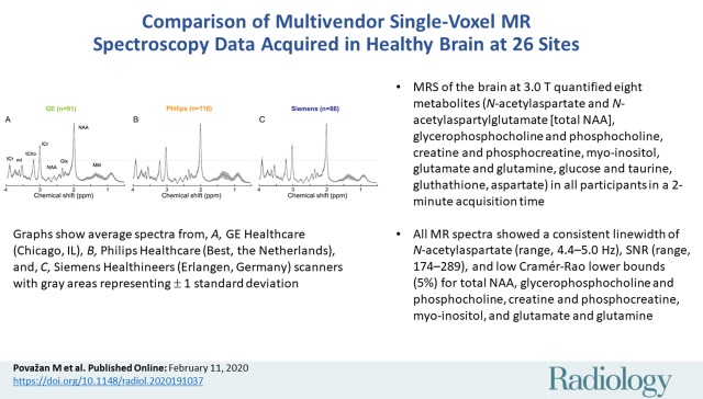
Background The hardware and software differences between MR vendors and individual sites influence the quantification of MR spectroscopy data. An analysis of a large data set may help to better understand sources of the total variance in quantified metabolite levels. Purpose To compare multisite quantitative brain MR spectroscopy data acquired in healthy participants at 26 sites by using the vendor-supplied single-voxel point-resolved spectroscopy (PRESS) sequence. Materials and Methods An MR spectroscopy protocol to acquire short-echo-time PRESS data from the midparietal region of the brain was disseminated to 26 research sites operating 3.0-T MR scanners from three different vendors. In this prospective study, healthy participants were scanned between July 2016 and December 2017. Data were analyzed by using software with simulated basis sets customized for each vendor implementation. The proportion of total variance attributed to vendor-, site-, and participant-related effects was estimated by using a linear mixed-effects model. values were derived through parametric bootstrapping of the linear mixed-effects models (denoted ). Results In total, 296 participants (mean age, 26 years ± 4.6; 155 women and 141 men) were scanned. Good-quality data were recorded from all sites, as evidenced by a consistent linewidth of -acetylaspartate (range, 4.4-5.0 Hz), signal-to-noise ratio (range, 174-289), and low Cramér-Rao lower bounds (≤5%) for all of the major metabolites. Among the major metabolites, no vendor effects were found for levels of myo-inositol ( > .90), -acetylaspartate and -acetylaspartylglutamate ( = .13), or glutamate and glutamine ( = .11). Among the smaller resonances, no vendor effects were found for ascorbate ( = .08), aspartate ( > .90), glutathione ( > .90), or lactate ( = .28). Conclusion Multisite multivendor single-voxel MR spectroscopy studies performed at 3.0 T can yield results that are coherent across vendors, provided that vendor differences in pulse sequence implementation are accounted for in data analysis. However, the site-related effects on variability were more profound and suggest the need for further standardization of spectroscopic protocols. © RSNA, 2020
背景 磁共振(MR)设备供应商和各检查部位之间的硬件和软件差异会影响磁共振波谱数据分析的结果。对大型数据集进行分析有助于更好地了解量化代谢物水平总变异性的来源。
目的 比较 26 个部位在健康志愿者中采集的多部位定量脑磁共振波谱数据,这些数据是使用供应商提供的单体素点分辨波谱(PRESS)序列采集的。
材料与方法 2016 年 7 月至 2017 年 12 月,这项前瞻性研究对来自 3 家不同供应商的 3.0-T MR 扫描仪的 26 个研究部位的脑中部顶区进行了短回波时间 PRESS 数据采集。使用针对每个供应商实施情况定制的模拟基础集软件对数据进行分析。采用线性混合效应模型来估计总方差归因于供应商、部位和参与者相关效应的比例。通过对线性混合效应模型进行参数自举(表示为 )来获得 值。
结果 共对 296 名志愿者(平均年龄,26 岁±4.6;155 名女性,141 名男性)进行了扫描。所有部位均记录到高质量的数据,这表现在 -N- 乙酰天冬氨酸(NAA)的线宽一致(范围,4.4-5.0 Hz)、信噪比(范围,174-289)和各主要代谢物的克里格下限低(≤5%)。在主要代谢物中,各代谢物的水平未见因供应商而产生差异,包括肌醇( >.90)、NAA 和 N- 乙酰天门冬氨酸谷氨酸( =.13)或谷氨酸和谷氨酰胺( =.11)。在较小的共振中,未见供应商对抗坏血酸( =.08)、天冬氨酸( >.90)、谷胱甘肽( >.90)或乳酸( =.28)产生影响。
结论 在 3.0 T 下进行多部位多供应商的单体素磁共振波谱研究,如果在数据分析中考虑到脉冲序列实施中的供应商差异,则结果在供应商之间是一致的。然而,部位相关的变异性影响更为显著,这表明需要进一步规范波谱协议。